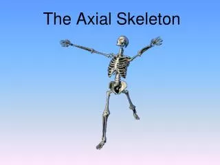
The Axial Skeleton
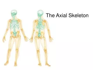
The Axial Skeleton. Sports Physiology Mr. Kottenstette. The Axial Skeleton. Forms the longitudinal axis of the body Divided into three parts Skull Vertebral column Bony thorax. The Axial Skeleton. Figure 5.6a. The Axial Skeleton. Figure 5.6b. The Skull. Two sets of bones Cranium

The Axial Skeleton
E N D
Presentation Transcript
The Axial Skeleton Sports Physiology Mr. Kottenstette
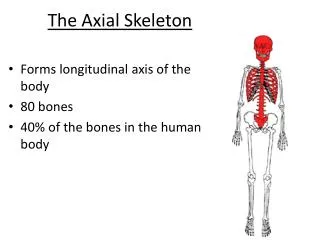
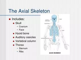
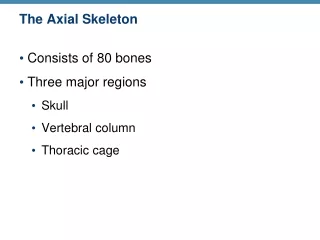
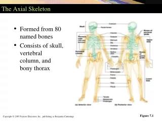
The Axial Skeleton • Forms the longitudinal axis of the body • Divided into three parts • Skull • Vertebral column • Bony thorax
The Axial Skeleton Figure 5.6a
The Axial Skeleton Figure 5.6b
The Skull • Two sets of bones • Cranium • Facial bones • Bones are joined by sutures • Only the mandible is attached by a freely movable joint
The Skull 28 named bones – 100’s of named features on the bones Important for muscular attachment Route for nerves, blood vessels
The Fetal Skull • The fetal skull is large compared to the infant’s total body length • Fontanels—fibrous membranes connecting the cranial bones • Allow the brain to grow • Convert to bone within 24 months after birth
The Hyoid Bone • The only bone that does not articulate with another bone • Serves as a moveable base for the tongue • Aids in swallowing and speech
The Vertebral Column • Each vertebrae is given a name according to its location • There are 24 single vertebral bones separated by intervertebral discs • Seven cervical vertebrae are in the neck • Twelve thoracic vertebrae are in the chest region • Five lumbar vertebrae are associated with the lower back
The Vertebral Column • Nine vertebrae fuse to form two composite bones • Sacrum • Coccyx
The Vertebral Column • Most adults have 26 total bones • 5 groups • Cervical - 7 • Thoracic - 12 • Lumbar - 5 • Sacral – 5, fused into single unit = Sacrum • Coccygeal – 4, fused into single unit = Coccyx • Form normal “S” curvature • Abnormal
The Vertebral Column • The spine has a normal curvature • Primary curvatures are the spinal curvatures of the thoracic and sacral regions • Present from birth • Secondary curvatures are the spinal curvatures of the cervical and lumbar regions • Develop after birth
Parts of a Vertebra • Body • Arch • Pedicle – stem/stalk • Lamina – roof • Transverse Processes • Spinous Process • Articular Facets • Joints
Atlas + Axis • Atlas = C1 • Articulates w/Skull = Atlantooccipotal joint • YES • Axis = C2 • Articulates w/Atlas via “Dens” = Atlantoaxial joint • NO
Unique Features – Cervical • Cervical C3-C7 • Transverse Foramen • Bifid Spinous Process • Horizontal Articular Facets
Unique Features - Thoracic • Thoracic T1-T12 • Spinous processes • Posterior Facing Facets • Multiple facets
Unique Features - Lumbar • Lumbar L1-L5 • Body • Medial/Lateral Facing Facets
Vertebral Canal - Home of the Spinal Cord • Foramen Magnum → L2 + Conus Medularis • Each vertebra in the C,T,L-spine has associated nerve • Intervertebral Foramen • T,L–Spine, nerve exits below • C-Spine, nerve exits above • Except… • Cauda Equina • Nerves exiting below L2 • Conus Medularis
Vertebral Ligaments • Anterior Longitudinal Ligament • Attached to anterior aspect vertebral bodies • ≥ ½ enclosure anterior aspect of column • Role
Vertebral Ligaments • Posterior Longitudinal Ligament • Attached to posterior aspect vertebral bodies, INSIDE vertebral canal • ≤¼ enclosure posterior aspect of column • Role
Vertebral Discs – Jelly Doughnut Annulus Fibrosis Nucleus Pulposis Herniation Affected nerve Most Common Level
Normal Sagittal Plane X-Section 1. Body of thoracic vertebra 2. Intervertebral disc 3. Spinal cord 4. Vertebral canal with spinal meninges 5. Spinous process of vertebra 6. Hyaline cartilage over articular surfaces of vertebral bodies 7. Anterior longitudinal ligament 8. Posterior longitudinal ligament
Sacrum and Coccyx • Sacrum • Formed by the fusion of five vertebrae • Coccyx • Formed from the fusion of three to five vertebrae • “Tailbone,” or remnant of a tail that other vertebrates have
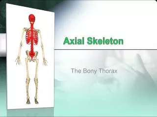
The Bony Thorax • Forms a cage to protect major organs • Consists of three parts • Sternum • Ribs • True ribs (pairs 1–7) • False ribs (pairs 8–12) • Floating ribs (pairs 11–12) • Thoracic vertebrae