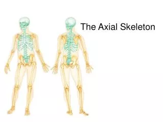
The Axial Skeleton
The Axial Skeleton. Forms longitudinal axis of the body 80 bones 40% of the bones in the human body. Axial Skeleton. Three Regions: Skull (8 cranial & 14 facial) ** bones associated with skull (6 auditory ossicles and hyoid) 2. Vertebral column (24 vertebrae, the sacrum & coccyx)

The Axial Skeleton
E N D
Presentation Transcript
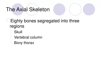
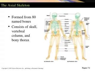
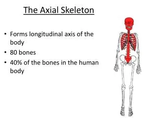
The Axial Skeleton • Forms longitudinal axis of the body • 80 bones • 40% of the bones in the human body
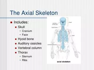
Axial Skeleton • Three Regions: • Skull (8 cranial & 14 facial) ** bones associated with skull (6 auditory ossicles and hyoid) 2. Vertebral column (24 vertebrae, the sacrum & coccyx) 3. Thoracic cage (sternum & 24 ribs)
The Skull • The bones of the skull protect the brain and guard the entrances to the digestive & respiratory systems • The skull (22 bones), the body’s most complex bony structure, is formed by the cranium (8 bones) and facial bones (14 bones) • 6 auditory ossicles (tiny bones) are situated within the temporal bones of the cranium (smallest bones in the body that are contained in the middle ear space; hammer, anvil, stirrup) • Hyoid bone (connected to the inferior surfaces of the temporal bones)
The Skull • Cranium – protects the brain and is the site of attachment for head and neck muscles • Facial bones • Supply the framework of the face, the sense organs, and the teeth • Provide openings for the passage of air and food • Anchor the facial muscles of expression
Anatomy of the Cranium Eight cranial bones: • 2 parietal • 2 temporal • Frontal • Occipital • Sphenoid • Ethmoid • The cranial bones enclose the cranial cavity, a fluid-filled chamber that cushions and supports the brain • Cranial bones are thin and remarkably strong for their weight
Skull – Anterior View Figure 7.2a
Frontal Bone • Forms the anterior portion of the cranium & the roof of the orbits (eye sockets)
Parietal Bones • Forms most of the superior and lateral aspects of the skull Figure 7.3a
Parietal Bones & Major Associated Sutures • Four sutures mark the articulations of the parietal bones 1.Coronal suture – articulation between parietal bones and frontal bone anteriorly 2. Sagittal suture – where right and left parietal bones meet superiorly
Parietal Bones & Major Associated Sutures 3. Lambdoid suture – where parietal bones meet the occipital bone (posterior) • 4. Squamosal or squamous suture – where parietal and temporal bones meet
Occipital Bone • Located at the back and lower part of the cranium
Temporal Bones Form part of both the lateral walls of the cranium & zygomatic arches Divided into four major regions: A. Squamous, B. Tympanic, C. Mastoid D. Petrous Figure 7.5
Sphenoid Bone • Butterfly-shaped bone that forms part of the floor of the cranium, unites the cranial and facial bones, and acts as a cross brace that strengthens the sides of the skull • Forms the central wedge that articulates with all other cranial bones
Ethmoid Bone • Most deep of the skull bones; lies between the sphenoid and nasal bones Figure 7.7
Facial Bones • Fourteen bones of which only the mandible and vomer are unpaired • The paired bones are the maxillae, zygomatics, nasals, lacrimals, palatines, and inferior conchae
Mandible • The mandible (lower jawbone) is the strongest bone of the face Figure 7.8a
Maxillary Bones • Medially fused bones that make up the upper jaw and the central portion of the facial skeleton (largest facial bones) Figure 7.8b
Zygomatic Bones • Irregularly shaped bones (cheekbones) that form the prominences of the cheeks and the inferolateral margins of the orbits
Other Facial Bones • Nasal bones – thin medially fused bones that form the bridge of the nose • Lacrimal bones – contribute to the medial walls of the orbit and contain a deep groove that house the tear ducts • Palatine bones– two bone plates that form portions of the hard palate and contribute to the floor of each orbit
Other Facial Bones • Vomer – forms part of the nasal septum • Inferior nasal conchae – paired, curved bones in the nasal cavity that form part of the lateral walls of the nasal cavity
Paranasal Sinuses Figure 7.11
Hyoid Bone • Lies just inferior to the mandible in the anterior neck • Only bone of the body that does not articulate directly with another bone • Attachment point for neck muscles that raise and lower the larynx during swallowing and speech Figure 7.12
Vertebral Column • 26 irregular bones (vertebrae) • Provide a column of support, bearing the weight of the head, neck, and trunk. • Transfers weight to the appendicular skeleton of the lower limbs • Protects spinal cord • Helps maintain an upright body position • Approx. length of an adult column is 71cm
Vertebral Column Cervical vertebrae 7 bones of the neck Thoracic vertebrae 12 bones of the torso Lumbar vertebrae 5 bones of the lower back Figure 7.13
Vertebral Column Sacrum - 5 fused vertebrae • bone inferior to the lumbar • vertebrae that articulates with the hip bones Coccyx – 4 fused vertebrae Figure 7.13
Disks are small shock absorbers between the vertebrae (gel-like interior)
General Structure of Vertebrae: • Vertebral body (centrum) – disc-shaped, weight-bearing region • Vertebral arch – composed of pedicles (walls) and flat layers called laminae (roof) ** forms the posterior margin of each vertebral foramen (together they form the vertebral canal which encloses the spinal cord) 3. Articular processes– projections on each vertebra
Cervical Vertebrae Table 7.2
Cervical Vertebrae • Most mammals have 7 cervical vertebrae (giraffes, whales, mice & humans) • Seven vertebrae (C1-C7) are the smallest, lightest vertebrae
Cervical Vertebrae: The Atlas (C1) • Holds up the head • The superior surface articulates with the occipital condyles of the skull (permits you to nod) • Has no body and no spinous process
Cervical Vertebrae: The Axis (C2) • The axis has a body, spine, and vertebral arches as do other cervical vertebrae • Articulates with the atlas to permit rotation Figure 7.16c
Thoracic Vertebrae • There are twelve vertebrae (T1-T12) • Distinctive heart-shaped body (more massive than that of a cervical vertebra) • Each thoracic vertebra articulate with ribs
Lumbar Vertebrae • The five lumbar vertebrae (L1-L5) are located in the small of the back and have an enhanced weight-bearing function • Largest vertebrae
Tip: Mealtimes Breakfast: 7 a.m. (7 cervical) Lunch: 12 p.m. (12 thoracic) Dinner: 5 p.m. (5 lumbar)
Sacrum and Coccyx • The sacrum • Consists of five fused vertebrae (S1-S5), which shape the posterior wall of the pelvis • Begin fusing after puberty and are completely fused at age 25-30 • Protects reproductive, digestive, and urinary organs • It articulates with L5 superiorly, and with the auricular surfaces of the hip bones
Coccyx • Coccyx (Tailbone) • The coccyx is made up of four (in some cases three to five) fused vertebrae that articulate superiorly with the sacrum • Generally begun fusing by age 26
Sacrum and Coccyx Figure 7.18a
Sacrum and Coccyx Figure 7.18b
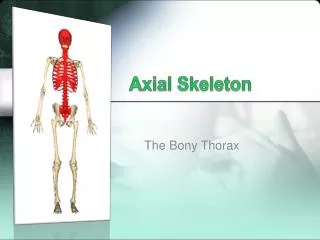
Bony Thorax (Thoracic Cage) • The thoracic cage is composed of the thoracic vertebrae, the ribs, and the sternum • Functions • Forms a protective cage around the heart, lungs, and great blood vessels • Supports the shoulder girdles and upper limbs • Provides attachment for many neck, back, chest, and shoulder muscles
Bony Thorax (Thoracic Cage) Figure 7.19a
Sternum (Breastbone) • A dagger-shaped, flat bone that lies in the anterior midline of the thorax • Fusion is not complete until at least age 25 (until this age the sternal body consist of four separate bones)
Ribs • There are twelve pair of ribs • All ribs attach posteriorly to the thoracic vertebrae • The superior 7 pair (true, or vertebrosternal ribs) attach directly to the sternum via costal cartilages • Ribs 8-10 (false, or vertebrocondral ribs) attach indirectly to the sternum via costal cartilage • Ribs 11-12 (floating, or vertebral ribs) have no anterior attachment
Ribs Figure 7.19a