The Axial Skeleton
The Axial Skeleton. I highly recommend Professor Wissman’s sites. For bones: http://homepage.smc.edu/wissmann_paul/bones/EBbonestutorial.html Check out all his links: http://homepage.smc.edu/wissmann_paul/anatomy1/ Also check out: Site for xrays & other diagnostic procedures:

The Axial Skeleton
E N D
Presentation Transcript
I highly recommend Professor Wissman’s sites • For bones: http://homepage.smc.edu/wissmann_paul/bones/EBbonestutorial.html • Check out all his links: http://homepage.smc.edu/wissmann_paul/anatomy1/ Also check out: • Site for xrays & other diagnostic procedures: http://www.radiologyinfo.org/en/sitemap/category.cfm?category=diag
http://homepage.smc.edu/wissmann_paul/bones/EBbonestutorial.htmlhttp://homepage.smc.edu/wissmann_paul/bones/EBbonestutorial.html This is an example of Prof Wissman’s bone site; this doesn’t show the roll-over answers
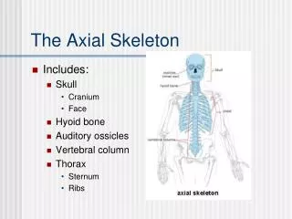
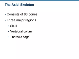
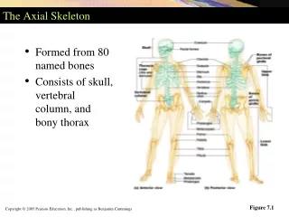
THE SKELETAL SYSTEMThe Axial Skeleton • The skeleton consists of • Bones (206) • Cartilages • Joints – also called articulations, are the junctions between skeletal elements • Ligaments – connect bones • Divided into axial and appendicular
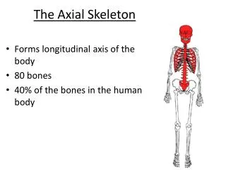
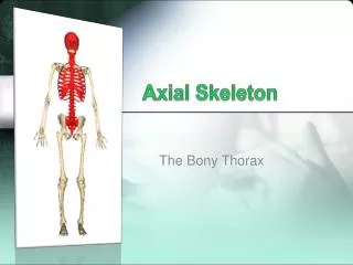
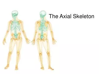
Axial skeleton - forms long axis of body • Skull • Vertebral column • Thoracic cage • Appendicular skeleton – appendages and what they attach to • Upper limbs (arms) • Pectoral girdle (shoulder) • Lower limbs (legs) • Pelvic girdle
Axial skeleton Skull Vertebral column Thoracic cage Axial skeleton is shown in green
The Skull • Cranial bones (or cranium) • Enclose the cranial cavity, which supports and protects the brain • Attachment sites for some head and neck muscles • Facial bones (anterior aspect of skull) • Form framework of face • Form cavities for sense organs of sight, taste and smell • Provides openings for passage of air and food • Hold the teeth • Anchor the muscles of the face
Cranium • Vault – “calvaria” = skullcap • Forms superior, lateral and posterior aspects of skull, and forehead • Base or floor: inferior part • Prominent bony ridges divide cranial base into 3 “fossae” (steps) – anterior, middle and posterior Anterior cranial fossa Middle cranial fossa Posterior cranial fossa (looking down on the floor of the skull)
Cranial bones • Frontal bone • Parietal bones (paired) • Occipital bone • Temporal bones (paired) • Sphenoid bone • Ethmoid bone
Cranial bones frontal parietal parietal parietal _______sphenoid temporal _____ethmoid occipital occipital
Temporal bones this is the right temporal bone looking at it from the right side
Ethmoid Small cranial bones… Sphenoid
Sutures • Immovable, interlocking joints of flat bones of skull • Irregular, saw-toothed appearance • Largest 4 skull sutures: where bones articulate with parietal bones • Coronal • Sagittal • Squamous • Lambdoid (FIND THEM)
Cranial “cavity” – houses brain • Smaller cavities • Housing middle and inner ear • Nasal cavity • Orbits • Sinuses • Openings (foramina, canals, fissures) for: • Spinal cord • Blood vessels • Twelve cranial nerves: I-XII
Remember, the skull is composed of: • Cranial bones (or cranium) [these were just reviewed] and • Facial bones (anterior aspect of skull) • Form framework of face • Form cavities for sense organs of sight, taste and smell • Provides openings for passage of air and food • Hold the teeth • Anchor the muscles of the face
Facial bones • Mandible • Vomer • Maxillae (paired) • Zygomatics (paired) • Nasal (paired) • Lacrimal (paired) • Palatines (paired) • Inferior nasal conchae (paired)
Facial bones: Mandible Vomer Maxillae (paired) Zygomatics (paired) Nasal (paired) Lacrimal (paired) Palatines (paired) Inferior nasal conchae (paired)
Maxilla (there are 2 which fuse, forming the upper jaw) Mandible (lower jaw)
(part of slide 18) Nasal cavity nasal bone ethmoid • Of bone and cartilage • Roof is ethmoid’s cribriform plate • Floor formed by palatine processes of the 2 maxillae and horizontal plates of palatine bones • These nasal-floor structures form roof of the mouth, called the hard palate inf nasal concha vomer maxilla___________
Nasal cavity To left, bones forming the left lateral wall of the nasal cavity (nasal septum removed) To right, nasal cavity with nasal septum in place, showing how the ethmoid bone, septal cartilage, and vomer make up the septum
Orbit Cone-shaped bony cavities holding the eyes, muscles that move the eyes, some fat and tear-producing glands; you don’t need to know all these bones that form it, just realize how complex it is and recognize the optic canal (optic nerve passes out through it) (right orbit shown)
Paranasal sinuses • Air-filled sacs in the bones • “Paranasal” because they cluster around and connect to the nasal cavity
Hyoid bone • Only bone which does not articulate with any other bone • Moveable base for the tongue • Points of attachment for neck muscles that raise and lower the larynx during swallowing
Remember that the Axial skeleton includes: Skull Vertebral column Thoracic cage Axial skeleton is shown in green
The Vertebral Column • Fetus and infant: 33 separate bones, or vertebrae • Adult: 24 vertebrae • Inferior 9 have fused forming • The sacrum (5) and • The coccyx (4)
Vertebrae • Cervical – 7 • Thoracic - 12 • Lumbar - 5 • Sacrum (5 fused) • Coccyx (4 fused)
Spinal curvatures • Cervical and lumbar are concave posteriorly* (lordosis) • Thoracic and sacral are convex posteriorly* (kyphosis) • Abnormal (see lab book p120): • Too much of either • Scoliosis (more than 10 degrees of lateral curvature) *when viewed from the side
Non-bony parts • Intervertebral discs • anulus fibrosis and nucleus pulposus) • Anterior longitudinal ligament • Posterior longitudinal ligament • Ligamentum flavum
Anterior longitudinal ligament: wide, strong and attaches to vertebrae as well as discs (prevents hyperextension) Posterior longitudinal ligament: narrow and relatively weak, attaching only to discs * Note “intervertebral foramen” vs “vertebral foramen” on next slides
Cervical vertebrae (C1-C7) C1 (atlas) C2 (axis)
Cervical Vertebrae • Smallest • Lightest • Most flexible • Triangular vertebral foramen • Transverse processes have foramina (transverse foramen) • Spinous process bifid (forked) except for C7
Thoracic Vertebrae T1-T12 • Heart shaped body • Additional small costal facets (costal=ribs) • Round or oval vertebral foramen • Form posterior part of rib cage
Lumbar Vertebrae L1-L5 • Massive blocklike bodies • Short, thick hatchet-shaped spinous processes • Limited mobility
Shapes posterior wall of pelvis Composite bone of 5 fused vertebrae Sacral foramina allow passage of vessels & nerves The Sacrum Coccyx (the tailbone)
Remember that the Axial skeleton includes: Skull Vertebral column Thoracic cage Axial skeleton is shown in green
Manubrium Body Xiphoid process True ribs 1-7 False ribs 8-12 Floating ribs 11,12 Sternum Ribs