The Axial Skeleton
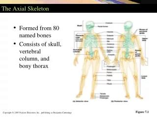
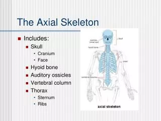
The Axial Skeleton. Includes: Skull Cranium Face Hyoid bone Auditory ossicles Vertebral column Thorax Sternum Ribs. Axial Skeleton: The Skull. The Skull. Contains 22 bones 8 Cranial Bones that enclose and protect the brain 14 Facial Bones that form the face. Cranial Bones.

The Axial Skeleton
E N D
Presentation Transcript
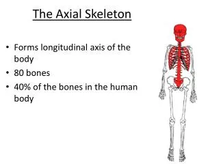
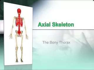
The Axial Skeleton • Includes: • Skull • Cranium • Face • Hyoid bone • Auditory ossicles • Vertebral column • Thorax • Sternum • Ribs
The Skull • Contains 22 bones • 8 Cranial Bones that enclose and protect the brain • 14 Facial Bones that form the face
Frontal Bone • Forms forehead, roofs of the eye sockets (orbits), and most of the front part of the cranial floor • Frontal sinuses lie deep within the bone
Parietal Bones • Form the sides and roof of the cranial cavity • Separated on top of skull by the sagittal suture
Temporal Bones • Form lower sides of the cranium and part of the cranial floor • External auditory meatus (ear canal) is located within these bones
Mastoid process is behind external auditory meatus and is a point of neck muscle attachment • Styloid process is point of neck and tongue muscle and ligament attachment • Mandibular fossa forms half of the temporomandibular joint with the mandible (lower jaw bone)
Occipital Bone • Forms posterior part and most of the cranial base • Foramen magnum passes through this bone • Occipital condyles are on either side of the foramen magnum that connect with the first vertebrae
Sphenoid Bone • The “keystone” of the cranial floor because it holds together all of the other cranial bones • The hypophyseal fossa is a depression for the pituitary gland
Ethmoid Bone • Light spongy bone in the anterior part of cranial floor between the eye sockets • Houses the nasal cavity • Contains the nasal conchae that cause turbulence in inhaled air, cleaning the air before it passes into the rest of the respiratory tract
Nasal Bones • Paired to form the bridge of the nose • The rest of the nose consists of cartilage
Maxillae • Paried to form the upper jawbone • Articulates with every bone in the face except the mandible • Forms the anterior 3/4 of the hard palate
Palatine Bones • Paired L-shaped bones • Form the posterior portion of the hard palate, part of the floor and lateral wall of the nasal cavity, and a small portion of the eye sockets
Mandible • The lower jawbone • Largest and strongest facial bone and only movable skull bone • Condylar process articulates with the mandibular fossa of the temporal bone to form the TMJ
Zygomatic Bones • Two cheekbones • Form the prominences of the cheeks and part of the lateral wall and floor of the eye sockets
Lacrimal Bones • Paired smallest bones of the face • Found near the tear ducts
Inferior Nasal Conchae • Scroll-like bones that project into the nasal cavity • Are below the ethmoid bone and other conchae
Vomer • Triangular bone on the floor of the nasal cavity • One of the parts of the nasal septum
Cleft Palate • Occurs when fusion of the left and right maxillary bones is not completed before birth • Repaired between 12 and 18 months with surgery
TMJ Syndrome • Caused by improperly aligned teeth, grinding or clenching teeth, trauma, or arthritis • Generally results in pain around the ear and jaw muscles
Deviated Nasal Septum • Nasal septum bends sideways from the middle of the nose • Can entirely block nasal passage in extreme cases
Sutures • An immovable joint • Found only between skull bones • Hold skull bones together
Coronal Suture- Between the frontal bone and two parietal bones • Sagittal Suture- Between the two parietal bones
Lambdoid Suture- between the parietal bones and occipital bone • Squamous Suture- between the parietal bones and temporal bones
Paranasal Sinuses • Paired cavities near nasal cavity • Located in the frontal bone, sphenoid bone, ethmoid bone, and maxillae • Lined with mucous membranes
Fontanels • Membrane-filled spaces found between cranial bones in infants • Replaced with bone by intramembranous ossification and become sutures • “Soft Spot” on baby’s head
Hyoid Bone • Does not articulate with or attach to any other bone • Suspended from the styloid processes by ligaments and muscles • Located in the neck between the mandible and larynx
Vertebral Column • Also called the spine or backbone • Composed of vertebrae • Functions as strong flexible rod that can rotate and move in all directions • Encloses and protects spinal cord • Supports the skull • Point of attachment for ribs, pelvic girdle, and back muscles
Regions of the Vertebrae • 7 cervical vertebrae in the neck • 12 thoracic vertebrae • 5 lumbar vertebrae supporting the lower back • 1 sacrum (consists of 5 fused sacral vertebrae) • 1 coccyx (consists of 4 fused coccygeal vertebrae)
Intervertebral Discs • Lie in between the vertebrae from the 2nd cervical vertebrae to the sacrum • Form strong joints, permit movement, and absorb vertical shock
Vertebral Column Curvature • The spine curves like a snake • Cervical and lumbar curves are convex (bulging out anteriorly) • Thoracic and sacral curves are concave (bulge out posteriorly)
Body • Thick, disc-shaped front portion • The weight-bearing part of a vertebra
Vertebral Arch • Extends backwards from the body of the vertebra • Formed by two short, thick processes (pedicles) that unite with the flat parts of the arch (laminae), ending with a single sharp, slender projection (spinous process)
Spinal Cord Openings • The vertebral foramen is the space between the vertebral arch and body that contains the spinal cord • All of the vertebral foramen combined forms the vertebral canal • The intervertebral foramen is the opening between adjoining vertebrae on both sides of the column contains a single spinal nerve
Transverse Processes • Extend laterally on each side where the lamina and pedicle join
Spinous Process • Projects from the junction of the laminae • Combined with the two transverse processes, these three are points of attachment for muscles to the vertebral column
Articular Processes • Superior Articular Processes join with the vertebra right above them • Inferior Articular Processes join with the vertebra right below them • The articulating surfaces are called facets and are lined with hyaline cartilage
Vertebrae • Numbered in sequence from top to bottom in each region
Cervical Vertebrae • All have three foramina: one verteral foramen and two transverse foramina
Atlas (C1 Vertebra) • Supports the head • Does not have a body or spinous process • Upper surface contains the superior articular facets that articulate with the occipital bone (allows you to nod “yes”)
Axis (C2 Vertebra) • Does have a body and spinous process • The dens, a tooth-shaped process, projects up through the vertebral foramen of the atlas and serves as a pivot to allow you to shake your head “no”
Remaining Cervical Vertebrae • C3 - C6 all follow the normal anatomy of the typical vertebra • C7 is also called the vertebra prominens; it has a single, large spinous process that can be felt at the base of the neck
Thoracic Vertebrae (T1 - T12) • Much larger and stronger than cervical vertebrae • Have facets for articulating with the ribs, which limits movement of the vertebrae