Download
1 / 18
200 likes | 523 Vues
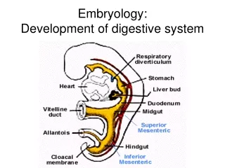
Embryology Digestive and Respiratory Systems. ANHB 2212 – Week 8 Avinash Bharadwaj. *. *. The Primitive Gut. Yolk Sac. Yolk Sac. Septum transversum. Note gut tube – foregut, midgut and hindgut Communication between midgut and yolk sac. The Definitive Gut.

E N D
EmbryologyDigestive and Respiratory Systems ANHB 2212 – Week 8 Avinash Bharadwaj
* * The Primitive Gut Yolk Sac Yolk Sac Septum transversum Note gut tube – foregut, midgut and hindgut Communication between midgut and yolk sac
The Definitive Gut Endodermal gut tube lining epithelium and glands only! Connective tissue and muscle is mesodermal. • Some features shown schematically! • Diaphragm • Abdominal foregut • Ventral and dorsal mesogastrium • Developing liver • Midgut • Elongates substantially • Vitellointestinal duct • Hindgut • Arteries from dorsal aorta
Further… • Ventral mesogastrium • Lesser omentum • Falciform ligament • Arteries • Coeliac • Foregut • Superior mesenteric • Midgut • Inferior mesenteric • Hindgut Lesser omentum Liver C Falciform Ligament SM IM
V D Abdominal Foregut • Abdominal part of oesophagus • Stomach • Ventral border – lesser curvature (attachment – lesser omentum) • Dorsal border – greater curvature (attachment – dorsal mesogastrium) • Proximal half of duodenum
Cranial V-I duct Caudal * Caecum The Midgut “Loop” • Superior mesenteric artery • Cranial (“Prearterial” ) limb (segment) • Vitellointestinal duct • “Postarterial” (caudal) limb • Caecum (Caecal “bud”) • Part of colon The coeliac and the inferior mesenteric arteries are omitted for clarity
C A V-I-D C C A V-I-D V-I-D C : caecum A : appendix V-I-D : vitellointestinal duct Rotation – Midgut Also note hindgut derivatives
Anal Canal • Separation from common cloaca • Partly ectodermal • “Proctodeum” • Anal membrane • Implications • Blood supply, venous drainage, nerves
Vitellointestinal Duct • Normally disappears • Persistence – Ileal (Meckel’s) diverticulum • Possibilities – ectopic mucosa (epithelium) • Proximity to the appendix • Partial persistence
R L Spleen Lesser Sac L.O. Liver F Stomach and Lesser Sac • Mesogastria • Divisions • Dorsal – splenic connections • Ventral – hepatic connections • Gastric borders
Lesser sac GS TC TMc C A GS V-I-D SI GS Peritoneal Cavity
Lesser sac P GS D X Peritoneal layers (mesothelium) X X TC TMc X X GS X Connective tissue SI GS Greater Omentum
V V D D Pancreas, Liver, Gall Bladder • Pancreas • Two “buds” • Differential duodenal growth • Fusion • Biliary tree stays with ventral duct • Hepatic and cystic parts liver and gall bladder • Liver – massive proliferation Blood supply : Liver and gall bladder – largely coeliac, S-m frequent Pancreas – always double
Examples of Anomalies • Hollow organs grow by proliferation of lining cells • Recanalisation by cell death essential • Failure of recanalisation atresia, stenosis • Oesophageal, duodenal, biliary • Anomalies of rotation • Pancreatic anomalies • Others • Imperforate anus • Pyloric stenosis (hypertrophic)
F H L M H Respiratory System • Offshoot of the digestive tube • Arises from the upper foregut • Single endodermal diverticulum • Ventral to the foregut • Branching
S Pt Coelom, Pleura, Lobes • Separation of pleural cavity • Note relationships • Phrenic nerve… • Lobes – right and left lungs
Lung – Histogenesis • Pseudoglandular – embryonic period • Canalicular : 16 – 26 weeks • Terminal sac phase : 7m Lung development determines the viability of a premature baby
Tracheo-oesophageal anomalies • Atresia – stenosis – fistula combinations.