Fractures and Skeletal Trauma
610 likes | 905 Vues
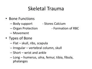
Fractures and Skeletal Trauma. Corinne Gratson, M.S., P.A.-C. Skeletal Trauma. Bone provides shape and support for the whole body It can be traumatized just like the soft tissues of the body 5 Basic Funcitons : -body support, organ protection, movement, calcium storage, hematopoiesis

Fractures and Skeletal Trauma
E N D
Presentation Transcript
Fractures and Skeletal Trauma Corinne Gratson, M.S., P.A.-C.
Skeletal Trauma • Bone provides shape and support for the whole body • It can be traumatized just like the soft tissues of the body • 5 Basic Funcitons: -body support, organ protection, movement, calcium storage, hematopoiesis • Types of Fractures: -Depressed, greenstick, impacted, longitudinal, spiral, oblique, transverse, comminuted, contrecoup, blowout, avulsion, stress, compound (open)
Types of Fractures Depressed
Greenstick Incomplete, more common in kids
Impacted Bone fragments are driven forcefully together
Longitudinal Follows the long axis of the bone
Spiral Bone is twisted
Oblique Fx runs oblique to axis of bone
Transverse Fx line @ 90° angle to long axis of bone
Comminuted Multiple fragements
Countrecoup Fx at site opposite site of impact (2° impact)
Avulsion Bony injury where a tendon or ligament attaches
Stress Low forces, repetitive for long period of time
Salter-Harris Fractures • Classification system used to diagnose fractures through or involving the physis (growth plate) in children • Types I-V
Salter-Harris • I – fracture across the growth plate (S – Slipped); • Tends to occur in younger kids; Xrays can look deceivingly normal
Salter-Harris • II – fracture starts across growth plate, then travels up the shaft, away from joint (A – Above) • Most common growth plate fx, older kids
Salter-Harris • III – starts through growth plate, then turns & exits into adjacent joint (L – Lower) • Joint cartilage gets disrupted; older kids
Salter-Harris • IV – starts above growth plate, goes through GP and into adjacent joint (Te – Through everything) • Disrupts joint cartilage; may need surgery; may impair proper growth
Salter-Harris • V – growth plate is crushed (R – Ruined/Rammed) • Most concerning as bone alignment & length can be affected
Salter-Harris Mnemonic • S (I) – Slipped (separation of physis) • A (II) – Above (metaphysis & physis) • L (III) – Lower (epiphysis & physis) • Te (IV) – Through everything (metaphysis, physis, epiphysis • R (V) – Ruined/Rammed (crushed physis)
What Happens When A Bone Breaks? • Fracture causes a break in the periosteum => disruption of local blood vessels & surrounding tissues => outpouring of blood and exudate (Inflammation Stage) • Blood & exudate => hematoma => fibrin in hematoma attempts to hold the fracture elements together
Components of Bone Formation • Cortex • Periosteum • Bone marrow • Soft tissue
Bone Healing • Fracture site begins to be remodeled with multiple types of cells (osteoblasts, osteocytes, osteoclasts), both from within the medullary canal and on the outside (periosteum) • This organization of cells helps to form a callus that surrounds the fracture site => matures into mineralized bone (Repair Stage) *chondrocytes produce Type II collagen; this cartilage is woven into bone by osteoblasts, eventually increasing Type I collagen
Bone Healing, cont. • Remodeling Stage • Woven bone is gradually converted to lamellar bone • Medullary cavity is reconstituted • Bone is restructured in response to stress and strain (Wolff’s Law)
How Does a Bone Heal? • In order for a bone to heal properly it must: • 1) Have an adequate blood supply • i.e., the less vascular compromise the better, whether it be from injuries, comorbidities or social hx (smoking) • 2) Have adequate mechanical stability
Blood Supply & Stabilty = Best Friends! • The earlier a fracture is stabilized, the better • Promotes revascularization • Substantial increase in blood flow to the fracture site (peaks 2 wks after fx) • After the 1st month interfragmentary movement increases callus formation
How Do We Treat Fractures? • Immobilize with a splint (making sure you have anatomic reduction with Xray assist.) • Helps reduce pain • Decrease damage to soft tissues, nerves, vascular supply (may even restore it after a reduction) • Decrease incidence of complications • Facilitate patient transport
Fx. Treatment, cont. • RICE! • Control pain & swelling (try to limit NSAIDs in case urgent surgery is necessary; stick to the narcs this time ) • NPO • If in hospital setting and surgery may be indicated or if pt. comes to your clinic and you plan to send pt. to ED
Fx Treatment, cont. • Complications of Prolonged Immobilization • Muscle atrophy • Other soft tissue atrophy • Loss of joint motion • Joint contractures (prolonged period of time) • Decrease in cartilage viability • Increased risk of VTE or PE
Fx. Treatment, cont. • Open Reduction Internal Fixation (ORIF) • Surgery with use of plates/screws (nails) to reduce the fracture • Provides rigid stabilization • Achieves anatomical or near-anatomical alignment • May allow for earlier weight bearing & ROM (depending on fx type, location & severity)
Fx. Treatment, cont. • Indications for ORIF • Fracture not reducible by closed method • Displaced intraarticularfx’s • Major avulsion fx’s • Nonunion fx’s that have not healed by means of closed reduction
Fx. Treatment, cont. • Disadvantages of ORIF • Cut on skin = increased risk for infection • Risk for neurovascular compromise • Hospitalization = $$$ • Risk for post-op scar tissue => ?limit range of motion? • Internal implants
Fx. Treatment, cont. • Comparing ORIF with Strict Immobilization • Depends on type, location & severity of fracture; also age of fx, comorbidities & age of pt. • Advantages of closed reduction include: • No increased risk for infection • No surgery • Likely no hospitalization
Fx. Treatment, cont. • Disadvantages of Closed Reduction & Immoblization • Near impossible to achieve anatomic alignment • If fracture is intraarticular (i.e., extends into the joint), pt. is at great risk for post-traumatic arthritis • May have lack of stability if ligamentous structures disrupted as well
Fx. Treatment, cont. • Other invasive treatment options include: • CRPP (Closed Reduction Percutaneous Pinning)
Fx. Treatment, cont. • External Fixator
Fx. Treatment, cont. • Ilizarov • Reduction, rigid fixation, early mobilization, early joint motion, functional loading and weight-bearing • For fx’s with severe soft tissue impairment, large bone loss, for intra- and peri-articularfx’s
Fractures in Real Life • Orthopaedic Consultation in ED • Is the pt. stable? What body part is fractured? Any other injuries? • Any concern for compartment syndrome? • Neurovascular status of affected extremity? • Open vs. Closed? • Open: Pt. needs tetanus shot, Abx (gram + coverage = 1st or 2nd Gen. Cephalosporin, poss. add gram neg. (aminoglycoside) coverage depending on severity of wound) x48hrs after fx & 48hrs after surgery; bedside I&D if possible; to OR within 6 hours
Is it or was it dislocated? • Will it need surgery? • Hip fxs, trimalleolarfxs, intraarticular distal radius, some acetabularfxs; some will be dependent upon amount of displacement
Compartment Syndrome • Limbs are divided into compartments of muscle, separated by fascia • Release of blood from the fracture site can cause an increase in interstitial pressure within the closed individual compartments • Swelling • Impaired venous and lymphatic return • Results in micro-vascular compromise
Compartment Syndrome • Causes • Trauma (fx) • Ischemic reperfusion following injury • Hemorrhage • Vascular punture • IVDU • Prolonged limb compression/crush injuries • Burns
Compartment Syndrome • The longer the pressure is there & the higher it gets, the more impaired is the myoneural function => soft tissue necrosis • Normal mean interstitial pressure is near 0 mmHg in non-contracting muscle • Pressure greater than 30 mmHg begins to compromise vessels & tissue causing nutrient flow, ischemia and pain • If diastolic BP exceeds compartment pressure by LESS than 30mmHg, this is considered an emergency (Whiteside’s Theory)
Compartment Syndrome • Diagnosis • Mostly a clinical diagnosis, but can also be tested by using a special needle to gauge the pressure (Stryker needle) • Pain*** earliest sign • Out of proportion to the injury • On passive stretch • Pallor • Paresthesias (early loss of vibratory sense) • Paralysis • Pulses (compare bilaterally) • **Once these occur, irreversible damage has occurred as well