Download
1 / 1
10 likes | 114 Vues
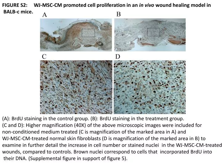
FIGURE S2: WJ -MSC-CM promoted cell proliferation in an in vivo wound healing model in BALB -c mice. (A): BrdU staining in the control group. (B): BrdU staining in the treatment group. ( C and D): Higher magnification (40X) of the above microscopic images were included for

E N D
FIGURE S2: WJ-MSC-CM promoted cell proliferation in an in vivo wound healing model in BALB-c mice. (A): BrdU staining in the control group. (B): BrdU staining in the treatment group. (C and D): Higher magnification (40X) of the above microscopic images were included for non-conditioned medium treated (C is magnification of the marked area in A) and WJ-MSC-CM-treated normal skin fibroblasts (D is magnification of the marked area in B) to examine in further detail the increase in cell number or stained nuclei in the WJ-MSC-CM-treated wounds, compared to controls. Brown nuclei correspond to cells that incorporated BrdUinto their DNA. (Supplemental figure in support of figure 5).