Understanding Cranial Nerve I and Its Role in Neuropsychology and Movement Disorders
This course module explores the significance of cranial nerve I (the olfactory nerve) in clinical neuropsychology, including its impact on disorders such as anosmia, Alzheimer’s, and Parkinson's disease. It provides detailed anatomical insights into the brain stem, thalamus, and basal ganglia, alongside their roles in common brain disorders. A specific focus will be on understanding movement disorders like Parkinson's and Huntington's disease, elucidating the neural circuitry involved in hypo- and hyperkinetic movements, and the implications for neuropsychological assessment and treatment.

Understanding Cranial Nerve I and Its Role in Neuropsychology and Movement Disorders
E N D
Presentation Transcript
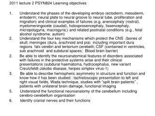
PSYN824 week 3 2011 Learning outcomes Understand importance of cranial nerve I for clinical neuropsychology practice Be able to identify structures on the dorsal and ventral brain stem and their involvement in common and uncommon brain disorders Describe the anatomy of the thalamus and the roles of the major nuclei Identify the structures that comprise the basal ganglia Describe the neuropsychology and neuropathology of Parkinson’s disease, Huntington’s disease Explain how disorders in basal ganglia circuitry produce hypo- and hyper-kinetic movements
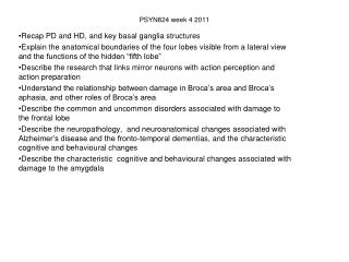
The cranial nerves Twelve pairs • labelled by Roman numerals, in a rostrocaudal sequence • Only the first two (Cranial nerves I and II) enter the CNS in the forebrain Three functions: • provide the sensory and motor information for the skin and muscles of the head and neck • convey to the cns the information from key sensory systems: vision, hearing, olfaction and taste 3. important components of the autonomic nervous system, important in visceral functions such as breathing, heart rate and blood pressure Cranial nerve 1 and neuropsychology • Can be damaged via cribiform plate leading to anosmia (olfactory loss) • Anosmia associated with frontal tumors, Alzheimer’s and possibly Parkinson’s disease • Olfactory nerves → olfactory bulb → olfactory tract → olfactory processing areas (broadly speaking the uncus, entorhinal and the peri- amygdaloid cortex) • Projectionds both ipsilaterally and contralaterally processing
Brain stem Brain stem: • dorsal aspect: inferior and superior colliculi, cerebellar peduncles and floor of fourth ventricle • ventral aspect: optic tract, mammillary bodies (and mammillo-thalamic tract) , infundibulum of hypothalamus (and pituitary stalk), cerebral peduncles, pons, pyramids, olives • not visible on surface : reticular formation (including the raphe n and locus coerulus) and the substantia nigra
What is the role of the mammillary bodies (MB) in memory ? Paired structure on brain stem Part of hypothalamus Integral part of limbic system Projects to anterior thalamus via mammillothalamic tract; principal input from hippocampus via post-commisural fornix Implicated in memory by patients with Wernicke-Korsakoff’s syndrome, single case and case series Kim et al (2009) report that left-sided only functional connectivity between MB and anterior thalamus significantly correlated with delayed verbal recall scores
Diencephalon: thalamus, hypothalamus, and mammillary bodies Thalamus: two halves are separated on the midline by the third ventricle but joined across the midline band of white tissue called the massa intermedia, landmark is the internal medullary lamina, a Y shaped sheet of white matter, it divides the grey matter into anterior, medial and lateral sections, contains the intralaminar n; also midline nuclei Intralaminar nuclei lesions are associated with cognitive deficits and typical “frontal like” behaviour Pulvinar: Wernicke’s aphasia Dorsomedial nucleus: anterograde amnesia Anterior thalamus/mammillary body: anterograde amnesia/frontal signs Lateral and medial geniculate nuclei are in the optic and auditory pathways Ventroposteromedial n and ventral posterolateral n involved in allodynia, parathesias Reticular nuclei: schizophrenia ? Persistent vegetative state ?
Persistent vegetative state Description not a prediction 50% reduction in cortical metabolism, which sometimes decreases over time No visual tracking, but orienting response, sleep wake cycle poor outcome predicted by: damage to thalamic (bilateral) diffuse regions of cortex, and to white matter regions May resolve to minimal state of consciousness
Owen et al (2006) reported a case where a 23 yo woman in VS for five month after TBI was able to “willfully modulate” her brain • Asked to imagine playing tennis (thinking of the ball volleying back and forth over the net) or walking through her own home, imaging what she could see • Increased activity in supplementary motor area (SMA), and parahippocampal gyrus (PPA), posterior parietal lobe (PPC) and lateral premotor cortex (PMC), respectively
From http://www9.biostr.washington.edu/da.html Note the relationship between caudate and putamen,
motor disorders linked to basal ganglia (aka “extrapyramidal” system) • diminished movements like in Parkinson’s disease • hyperkinetic disorders like Huntington’s disease, Tourette’s and ballism • motor stereotypies such as Leigh-Nyham syndrome (self mutilations) • OCD incl punding (of course, OCD is not a pure motor disorder)
Parkinson’s disease (paralysis agitans or shaking palsy) clinical triad of Parkinson’s disease (PD) is a progressive degenerative disorder which affects about 1% of the population over 65; The classical clinical motor triad is • bradykinesia and akinesia including “iron mask” of PD • muscular rigidity, in particular cogwheel rigidity • resting tremor Pathological hall marks • progressive loss of the dopaminergic neurons in the darkly pigmented substantia nigra pars compacta • presence of intraneuronal inclusions in the cell bodies called Lewy bodies in PD with dementia
Hoehn and Yahr Staging of Parkinson’s disease • Stage One: unilateral disease, minimal functional impairment • Stage Two: bilateral disease, without impairment of balance • Stage Three: mild to moderate bilateral disease, some postural instability, physically independent • Stage Four: severe disability, still able to walk or stand unassisted but markedly physically incapacitated • Stage Five: wheelchair bound or bedridden unless aided
Non-motor impairment in Parkinson’s disease 1. Cognitive impairment is independently associated with depression, sleep disturbance and anxiety • Depression 25% • Dementia 29%, lifetime risk 80% (Riedel et al 2010) • MCI present 19% at diagnosis Aarsland et al (2009) As a whole, the data show that, compared to demographic peers, in PD without dementia there are impairments in • Attention and executive function • Memory • Visuospatial functions • Language Presence of bradyphrenia 2. Disorders of impulse control
Abbreviations: SSC, somatosensory cortex; PMC, primary motor cortex; PreMotor, Premotor cortex; PFC, prefrontal cortex; PPC, posterior parietal cortex; DLPFC, dorsolateral prefrontal cortex; GPe, globus pallidus external; GPi, globus pallidus internal; MD, mediodorsal nucleus of the thalamus; VA, ventral anterior nucleus of the thalamus; VL, ventrolateral nucleus of the thalamus, SNr, substantia nigra pars reticulata; SNc, substantia nigra pars compacta; OFC, orbitofrontal cortexTable modified from from Blumenfeld 2010
Huntington’s disease • Inherited, neurodegenerative disease, autosomal dominant disorder, abnormal gene on chromosome 4 mapped in 1983 • Affects approximately 5-10/100,000 • Presents with triad of symptoms: motor, cognitive, psychiatric (independent) • Average age at onset 30-40 years • Average duration of disease onset until death 10-17 years • http://www.youtube.com/watch?v=AxnTSFPbY2A Sprengelmeyer et al (1996) reported that in preclinical patients there was impaired recognition of facial expression of disgust • Problems with replication ? • Substantial heterogeneity in clinical presentation • Volume of the anterior insula ?
Chiccetti et al (2011) Brain 134; 641–652 Figure 3 Cell implants performed in patients with Huntington’s disease and evaluated 18 months following transplantation showed good survival of the grafted neurons, despite the presence of some microglial activation surrounding the grafted areas. Cell implants evaluated 10 years after transplantation showed significant microglial activation specifically in striatal zones of the grafts, which also received cortical glutamatergic connections.
Movement disorders • Relative pattern of activity in direct and indirect pathways determines movement • Direct pathway “starts” movements • Indirect pathways involved in “stopping” and “switches between” movements • Neurons in the Gpi have high intrinsic rate of firing, need to be silenced to start movement, activated to stop movement