Unit 5: The Integumentary System
Unit 5: The Integumentary System. The Integumentary System. The skin and its derivatives (hair, nails, glands, ect…) which provide the external protective covering of the body. Functions. Protection : against abrasions, bacterial invasion, UV light, dehydration

Unit 5: The Integumentary System
E N D
Presentation Transcript
The Integumentary System The skin and its derivatives (hair, nails, glands, ect…) which provide the external protective covering of the body
Functions • Protection: against abrasions, bacterial invasion, UV light, dehydration • Stimulus perception: nerve endings in dermis detect: temp, pressure and pain • Excretion: perspiration releases body waste • Regulates body Temp: via perspiration • Synthesis of vitamin D: via exposure to UV light • Blood reservoir: dermis holds large volume of blood until needed elsewhere
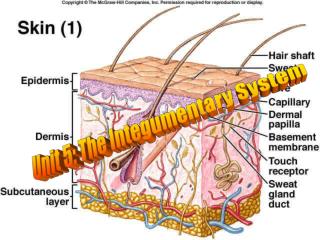
3 Layers • Epidermis: epithelial cells ( stratified squamous) • Dermis: dense irregular connective tissue • Hypodermis: areolar and adipose
Epidermis • Made up of : • stratified squamous epithelium • karatinocytes ( produce keratin) • melanocytes ( produce melanin for skin pigment) • langerhan’s cells and gransteid cells ( for immunity.
Sublayers of epidermis ( called strata) Starting with deepest • Stratum basale: mitotic layer, 1 cell thick • It continuously divides pushing the cells above them toward the surface, • they can change and slowly die before being shed • Stratum spinosum: 8-10 rows of irregularly shaped cells. • Begin producing keratine • Stratum granulosum: 3-4 rows of darkly stained cells. • cell nuclei begin to degenerate as layers receives less nourishment
Stratum lucidum: 2-3 rows of clearish cells only found in thick skin of palms and soles • Stratum corneum: 25 + rows of shingle-like cells remnants. The protective waterproof layer layer Epidermis: • No blood vessels • Nourished by diffusion ( does not make it to last 2 to 3 strata • Takes about 2 to 3 weeks to completely replace skin ( basale to corneum)
Dermis Thicker connective tissue layer beneath epidermis, contains collagen and elastic fibers
2 Regions of Dermis • Papillary region • Contains dermal papilla which are dermal projections into epidermis • Some papilla contain blood vessels and pain receptors or touch receptors (meissner’s corpuscles) • The dermal papilla produces ridges and valleys on the surface of epidermis ( increases friction for grip) • Sweat glands have glands that open at ridge peaks (making peaks sweaty) and reason for over fingerprints
2 Regions of Dermis • Reticular region • Contains: • Dense irregular connective tissue • Adipose tissue • Hair follicles • Oil and sweat glands • This region provides strength and elasticity to the skin • With age, elasticity lessens • Repeated stretched areas don’t bounce back… they wrinkle • IF overstretched (pregnancy) they tear leaving stretch marks (scars)
Hypodermis • Also called superficial fascia • Composed of aereolar and adipose connective tissue (usually muscle) • Acts as a shock absorber and insulator due to fat and water content • Contains Pacinian corpuscles (nerve endings for pressure) • Technically not part of integument
Skin color • Would be clear if not for 3 pigments: • Melanin: • Carotene • Hemoglobin
Melanin: • Range in color from orange to brown to dark brown, • All people have it except albinos • Amount produced is influenced by: genetics and exposure to UV light
Carotene • Yellowish pigment • Collects in stratum corneum and adipose
Hemoglobin • Oxygen carrying blood pigment • Gives skin its pinkness • So skin color is a blend of 3 pigments as influenced by genetics and environment
Epidermal Derivatives • Hair: somewhat protective ( nose, eyelashes or from sun/cold) • A hair (pili) consists of: • Shaft: visible portion • consisting of 3 layers of dead cells: 1. cuticle (outer) 2. medulla (inner) and 3. cortex (middle) • Root: portion of hair below surface, extends into the dermis and possible even hypodermis. Has the same 3 layers.
The root is surrounded by epidermal extensions which form the hair follicle ( sac around the hair) • The enlarged base of the root is called the bulb • The indention into the bulb is called the papilla of hair (contains blood vessels)
Epidermal Derivatives • Arrector pili muscle: • smooth muscle attached to the follicle and anchored in dermis • pulls air up straight out from skin in response to cold and fright
Nails • Another epidermal modification with little remaining function • Are hard keratinized dead cells produced by stratum basale, but nail itself is stratum corneum
Glands • Sebaceous glands: oil • Exocrine glands, usually associated with hair follicle • An oily mixture of cholesterol, lipids and cell fragments is called sebum • Sebum: protects hair from drying, becoming too brittle and from bacterial growth • A blocked sebaceous gland is a whitehead ( or a blackhead if the melanin and sebum oxidize), if it is infected it is a pimple
Glands • Sudoriferous gland: (sweat) • Sweat: mixture of water, salt and organic waste ( very similar to urine) • Functions: • eliminate waste • temp regulator
Types of sudoriferous glands • Eccrine: small coiled tubular glands release “sweat” • Found everywhere except lips, nipples and genital • Sweat is about 99% water • Apocrine:largely still tubular, but ducts open into hair follicles • Found only in axillary and pelvic regions and do not begin to function until stimulated by hormones at puberty • Produce thicker sweat with more organic wastes ( likely to have an odor due to bacteria living on it)
Ceruminous gland: modified apocrine sudoriferous glands • Found in external auditory meatus ear canal) • Secrete a fluid which reacts with sebum to produce cerumen ( a waxy protective substance)
Injury and Tissue Repair • Contact inhibition: • Cells will migrate and continue to divide until they come in contact with other cells of the same tissue type • A wound which only penetrates as far as stratum basale ( epidermis) • Heals with no scar tissue
Deep wound healing: • Inflammation: first response to damage • The damage cells release histamine (cause vascular dilation (bigger) and increase cell membrane permeability) • This causes more blood to enter wound ( swelling, heat and redness) • The increased blood delivered increase nutrition, and blood clotting factors and macrophages (WBC)
A blood clot forms scaling wound from both external world and surrounding healthy cells • The surviving cells next to the wound divide beneath the necrotic (dead) tissue repairing the wound • Since this new tissue does not match the original, it is called granulation tissue (scar tissue)
Regeneration: replacement of destroyed tissue by same kind of cells • Fibrosis: process of fibrous connective tissue repair ( scar) • Scab: blood clot and dead tissue which are sealed from healthy cells
So tissue repair depends on type of tissue damaged • Epithelial tissue: heals rapidly and nearly perfect • Connective tissue: repairs fairly rapid, but is imperfect (scars) • Muscles and nervous tissue: hardly replace the destroyed cell at all, just work around them
Burns • Tissue damage by heat, electricity, radioactivity or chemical causes • Burns destroy protective epidermis allowing: • Microbial infection • Extensive fluid, electrolyte, protein loss • Loss of temperature
Burns • Immediate danger of burns is the loss of fluid and electrolytes ( reduces volume of blood, renal shutdown and shock) • After fluid replacement the first 24 hrs, the major damage is infection • Burns are classified by depth and percentage of surface area affected:
Burn Classification • 1st degree: surface epidermis only ( no scarring) ex sunburn • 2nd degree: epidermis and some dermis, blistering and pain, mild scarring • 3rd degree: integumentary system or more is destroyed, no pain, charred dry appearance extensive scarring slow healing, skin graft or death
Cancer • Tumor or neoplasm: excessive growth of tissue uncontrolled cell division • Benign tumor: harmless, cells don’t spread (stay in one clump) • Malignant: cells keep dividing, spreading and invading other body areas • Metastasis: migration of cancer cells and other body parts. They produce 2ndary tumors and usually the cause of death in cancer
Sarcoma: general term for cancer of connective tissue • Adeosarcoma: cancer in a gland • Myeloma: cancer in bone marrow • Osteogenic sarcoma: bone cancer
Melanoma • Melanoma can grow very quickly. • It can become life-threatening in as little as six weeks and if untreated, it can spread to other parts of the body. • It can appear on skin not normally exposed to the sun. • It is usually flat with an uneven smudgy outline. • It may be blotchy and more than one color – brown, black, blue, red or grey.