Download

1 / 25
250 likes | 442 Vues
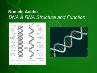
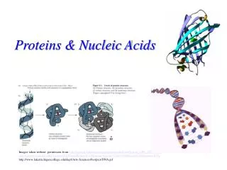
Complex Structure and Function of Proteins and Nucleic Acids. Monomers of Nucleic Acids are? Nucleotides…made up of? Nitrogenous base; sugar; and phosphate group. Monomers of proteins are? Amino Acids. Jobs that Proteins Do…. Substrate binds to enzyme. 2. 2.

E N D
Complex Structure and Function of Proteins and Nucleic Acids Monomers of Nucleic Acids are? Nucleotides…made up of? Nitrogenous base; sugar; and phosphate group Monomers of proteins are? Amino Acids
Jobs that Proteins Do… Substrate binds to enzyme. 2 2 1 Active site is available for a molecule of substrate, the reactant on which the enzyme acts. Substrate (sucrose) Glucose Enzyme (sucrase) OH H2O Fructose H O 3Substrate is converted to products. Enzymes • Are a type of protein that acts as a catalyst, speeding up chemical reactions • When bound to substrate, they form enzyme substrate complex Most importantly, while the substrate (sucrose) changes, the enzyme remains unchanged throughout the cycle! What was the name of the process that adds water, and breaks down a molecule? Hydrolysis!! 4Products are released.
Most chemical reactions that take place in the body, do so in the presence of enzymes. Why? Most chemical reactions would take a long time without enzymes. Our body cells produce toxins as waste products during cellular respiration, such as H2O2 (hydrogen peroxide). At high enough levels, H2O2 can be very toxic. The body has a response, however, by using an enzyme, which renders the H2O2 into less dangerous substances…H20 and 02 Using enzymes means less energy must go into the reaction! Any body process which conserves energy is good!
What does a catalyst do? Why are enzymes important? What are the subunits (or monomers) of proteins? What each of the following (A-E) in the diagram above? Which letter represents the activation energy required without the enzyme present? B A
Polypeptides • Polypeptides • Are polymers (long chains) of amino acids • A protein • Consists of one or more polypeptides • Amino acids • Are organic molecules possessing both carboxyl and amino groups • Differ in their properties due to differing side chains, called R groups
Amino acids • Are linked by peptide bonds A peptide bond is a chemical bond formed between two molecules when the carboxyl group of one molecule reacts with the amine group of the other molecule, thereby releasing a molecule of water (H2O). This is a dehydration synthesis reaction (also known as a condensation reaction), and usually occurs between amino acids. In what other biomolecules did we see dehydration synthesis occurring? Carbohydrates!
CH3 CH3 CH3 CH CH2 CH3 CH3 H CH3 H3C CH3 CH2 CH O O O O O H3N+ H3N+ H3N+ H3N+ C H3N+ C C C C C C C C C O– O– O– O– O– H H H H H Valine (Val) Leucine (Leu) Isoleucine (Ile) Glycine (Gly) Alanine (Ala) Nonpolar CH3 CH2 S H2C CH2 O NH CH2 H2N C C CH2 CH2 O– CH2 O O O H H3N+ H3N+ C C C C H3N+ C C O– O– O– H H H Phenylalanine (Phe) Proline (Pro) Methionine (Met) Tryptophan (Trp) Figure 5.17 the 20 Amino Acids and their properties...
OH NH2 O C NH2 O C OH SH CH2 CH3 OH Polar CH2 CH CH2 CH2 CH2 CH2 O O O O O O H3N+ C H3N+ C H3N+ C C H3N+ C C H3N+ C C C C C H3N+ C O– O– O– O– O– O– H H H H H H Glutamine (Gln) Tyrosine (Tyr) Asparagine (Asn) Cysteine (Cys) Serine (Ser) Threonine (Thr) Basic Acidic NH3+ NH2 NH+ O– O –O O CH2 C NH2+ C C NH Electrically charged CH2 CH2 CH2 CH2 CH2 O O CH2 CH2 C CH2 C H3N+ C H3N+ C O O– O– CH2 C H3N+ CH2 C H O H O– C C H3N+ CH2 H O O– C C H3N+ H O– H Lysine (Lys) Histidine (His) Arginine (Arg) Glutamic acid (Glu) Aspartic acid (Asp) the 20 Amino Acids...continued
What do peptide bonds help to form? What type of molecule do many polypeptides join together to make? Describe the structure of an amino acid. What differentiates each amino acid?
Amino acid subunits +H3NAmino end Pro Thr Gly Gly Thr Gly Glu Seu Lys Cys Pro Leu Met Val Lys Val Leu Asp Ala Arg Val Gly Ser Pro Ala Glu Lle Asp Thr Lys Ser Tyr Trp Lys Ala Leu Gly lle Ser Pro Phe His Glu His Ala Glu Val Thr Phe Val Ala Asn lle Thr Asp Ala Tyr Arg Ser Ala Arg Pro Gly Leu Leu Ser Pro Tyr Ser Tyr Ser Thr Thr Ala o Val c Val Glu – Lys o Thr Pro Asn Carboxyl end Protein Conformation and Function A protein’s specific conformation (shape) determines how it functions There are four levels of protein structure. • Primary structure • Is the unique sequence of amino acids in a polypeptide
H H H H H H O O O O O O O H H H H H H R R R R R R R C C C C C C C C C C C C C N N N N N N N N N N N N N C C C C C C C C C C C C C C R R R R R R H H H H H H H O O O O O O O H H H H H H H pleated sheet H O H H Amino acidsubunits C C N N N C C C R H O H H H H H H N N N N N N helix C C O C H H H C C C R R R R R H H C C C C C C O O O O H C R O C C O H C O N N H C C R R • Secondary structure • Is the folding or coiling of the polypeptide into a repeating configuration • Includes the helix and the pleated sheet Capture silk and drag lines are built of differing secondary structures, and has a similar structure to kevlar!
Hydrophobic interactions and van der Waalsinteractions CH CH2 CH2 H3C CH3 OH Polypeptidebackbone H3C CH3 Hyrdogenbond CH O HO C CH2 CH2 S S CH2 Disulfide bridge O -O C CH2 CH2 NH3+ Ionic bond • Tertiary structure • Is the overall three-dimensional shape of a polypeptide • Results from interactions between amino acids and R groups • Is most related to specificity of function
Polypeptidechain Collagen Chains Iron Heme Chains Hemoglobin • Quaternary structure • Is the overall protein structure that results from the aggregation, or fusion of two or more polypeptide subunits
Sickle-Cell Disease: A Simple Change in Primary Structure Normal hemoglobin Sickle-cell hemoglobin Primary structure Primary structure . . . . . . Exposed hydrophobic region Val His Leu Thr Pro Glul Glu Val His Leu Pro Glu Thr Val 5 6 7 3 4 5 6 7 1 2 1 2 3 4 Secondaryand tertiarystructures Secondaryand tertiarystructures subunit subunit Quaternary structure Hemoglobin A Quaternary structure Hemoglobin S Molecules interact with one another tocrystallize into a fiber, capacity to carry oxygen is greatly reduced. Function Molecules donot associatewith oneanother, eachcarries oxygen. Function 10 m 10 m Normal cells arefull of individualhemoglobinmolecules, eachcarrying oxygen Red bloodcell shape Red bloodcell shape Figure 5.21 • Results from a single amino acid substitution in the protein hemoglobin Fibers of abnormalhemoglobin deform cell into sickle shape.
Which of the following shows the primary structure of a protein? Which shows the secondary structure of a protein? Which shows the tertiary structure? Which of the protein structures is most responsible for the sickle-cell mutation? A B C
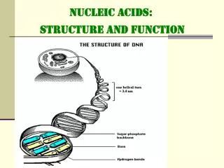
The Structure of Nucleic Acids • Nucleic acids • Exist as polymers called polynucleotides The phosphate group, with its slightly negative charge, makes the overall charge of DNA slightly negative as well. • Each polynucleotide • Consists of monomers called nucleotides • Sugar + phosphate + nitrogen base • Nucleotide monomers • Are made up of nucleosides (sugar + base) and phosphate groups • The portion of a nucleotide without the phosphate group is called a nucleoside
Nitrogenous bases Pyrimidines Structure of Nucleosides: Bases, and Sugars Cytosine (C) Thymine (T, in DNA) Uracil (U, in RNA) Purines Adenine (A) Guanine (G) Sugars Deoxyribose (in DNA) Ribose (in RNA)
Nucleotide Polymers • Nucleotide polymers are linked together to build a polynucleotide • Adjacent nucleotides are joined by covalent bonds that form between the –OH group on the 3 carbon of one nucleotide and the phosphate on the 5 carbon on the next • These links create a backbone of sugar-phosphate units with nitrogenous bases as appendages • The sequence of bases along a DNA or mRNA polymer is unique for each gene (which is a region on a DNA molecule)
5 O 3 3 O P P 5 5 C O G 1 3 2 4 4 2 1 3 5 O P P T A 3 5 O O 5 P P 3 DNA Double Helix When guanine and cytosine bond, they form triple hydrogen bonds H-bonds When thymine and adenine bond, a double hydrogen bond is formed
What is the structure of a nucleoside? What three subunits go into a nucleotide? In nitrogenous bases, what are the differences between purines and pyrimidines? What is the structural difference between ribose, and deoxyribose? What type of bond joins nucleotides together? What two subunits of a nucleotide form the backbone of the DNA molecule? What type of bond joins nitrogenous bases in the center of the DNA double helix?
DNA Structure The nucleotides connect at the hydroxyl group of the 5’ carbon sugar (at the 3’ end) • Antiparallel nature: • • Sometimes called complementary” sugar/phosphate backbone runs in opposite directions • one strand runs 5’ to 3’, while the other runs 3’ to 5’; One DNA molecule includes many genes
Scientists can use DNA and Proteins as Tape Measures of Evolution Molecular comparisons • Help biologists sort out the evolutionary connections among species • How similar are the sequences of nucleotides? • The closer the sequence, the closer the relationship • Remember, all life, from the simplest prokaryote to the most complex eukaryote, contains the same four nitrogenous bases. It is simply the sequence of base-pairs, and amount of DNA that differs from organism to organism!
ATP Adenosine triphosphate is a common source of activation energy for metabolic reactions. ATP is essentially an RNA adenine (adenosine) nucleotide with two additional phosphate groups. The wavy lines between these two phosphate groups indicate high energy bonds. When that last bond is broken, and the ATP is converted to ADP (adenosine diphosphate), energy is released, and can be used to spur a reaction. Conversely, a new ATP molecule can be built by combining ADP and a phosphate through a process known as phosphorylation using energy obtained from glucose.
Why is DNA’s structure sometimes referred to as “antiparallel”? How can scientists determine relatedness of species? How does ATP differ from RNA nucleotides of adenine? How does ATP provide energy to be used by the body? What process builds ATP molecules?