血液的主要功能 Function of Blood
血液的主要功能 Function of Blood. 夏 强, MD & PhD 浙江大学医学院生理学系 医学院科研楼 C 座 518 室 电话: 88208252 Email : xiaqiang@zju.edu.cn. 血液的成分. Plasma (血浆) Blood Cells Red Blood Cells (RBC) or Erythrocytes (红细胞) White Blood Cells (WBC) or Leucocytes (白细胞) Platelets (PLT) or Thrombocytes (血小板).

血液的主要功能 Function of Blood
E N D
Presentation Transcript
血液的主要功能Functionof Blood 夏 强,MD & PhD 浙江大学医学院生理学系 医学院科研楼C座518室 电话:88208252 Email:xiaqiang@zju.edu.cn
血液的成分 • Plasma(血浆) • Blood Cells • Red Blood Cells (RBC) or Erythrocytes(红细胞) • White Blood Cells (WBC) or Leucocytes(白细胞) • Platelets (PLT) or Thrombocytes(血小板)
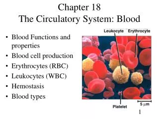
Hematocrit(packed cell volume, 血细胞比容) The hematocrit is a rapid assessment of blood composition.It is the percent of the blood volume that is composed of RBCs (red blood cells). Plasma includes water, ions, proteins, nutrients, hormones, wastes, etc.
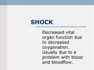
血量 • 是指全身血液的总量 • 占体重的7-8% 休克种类的图示。 没有休克 (左), 血管扩张,分布性休克 (中), 低血容量休克,失血引起 (右)
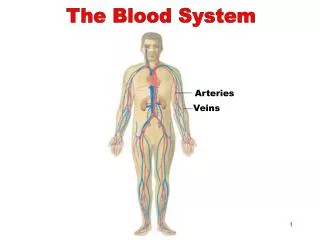
血液的功能 • 运输 • O2 and CO2 • Nutrients (glucose, lipids, amino acids) • Waste products (e.g., metabolites) • Hormones • 调节 • pH • Body temperature • 保护 • Blood coagulation • Immunity
红细胞Red blood cells (Erythrocytes) • RBC count • M: 4.0~5.5×10¹²/L • F: 3.5~5.0×10¹²/L • Hemoglobin(血红蛋白) • M: 120~160 g/L • F: 110~150 g/L • 功能: • 运输O2和CO2 • 缓冲
d Erythrocyte Sedimentation Rate (ESR)(红细胞沉降率) • The distance that red blood cells settle in a tube of blood in one hour • Normal value [Westergren method(魏氏法,国际血液学标准化委员会推荐魏氏法为标准法)]: M: 0~15 mm/h,F: 0~20 mm/h • An indication of inflammation which increases in many diseases, such as tuberculosis & rheumatoid arthritis… International Council for Standardization in Haematology (ICSH)
Iron deficiency anemia (缺铁性贫血)
Hemolysis(溶血) Red blood cells without (left and middle) and with (right) hemolysis. Note that the hemolyzed sample is transparent, because there are no cells to scatter light.
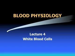
WBC count WBC Count (109/L) % Granulocytes粒细胞 Neutrophils中性粒细胞 2.0~7.0 50~70 Eosinophils嗜酸性粒细胞 0.02~0.5 0.5~5 Basophils嗜碱性粒细胞 0~0.1 0~1 Monocytes单核细胞 0.12~0.8 3~8 Lymphocytes淋巴细胞 0.8~4.0 20~40 Total 4~10
血小板Platelets (Thrombocytes) • 正常值: • (100~300) x 109/L • 主要参与止血
血小板的生理特性 1. 黏附Adhesion Platelets adhere to the vessel wall at the site of injury von Willebrand factor, vWF
Unifying model of platelet adhesion to collagen at arterial shear. Two different pathways by which human and mouse platelets firmly adhere to collagen at arterial shear are illustrated. In both, the majority of platelets are initially tethered to collagen via GP Ib/IX/V interacting with collagen-bound VWF (left), although a minority of platelets interact directly with collagen independently of VWF/GP Ib/IX/V. In the first pathway (upper), signaling from GP VI first leads to activation of integrins α2β1 (GP Ia/IIa) and αIIbβ3 (GP IIb/IIIa). Activated integrins then firmly attach the platelet to collagen, either directly (α2β1) or via collagen-bound VWF (αIIbβ3) (right). In the second pathway (lower), platelets first adhere to collagen via integrin α2β1, before GP VI engages collagen and induces activation. These two pathways are likely to reinforce each other and the events of thrombus formation. Release of secondary mediators (ADP and TxA2) would further potentiate these events (right). (Redrawn from Auger JM, Kuijpers MJ, Senis YA: Adhesion of human and mouse platelets to collagen under shear: a unifying model. FASEB J 2005;19:825-827.)
2. 聚集Aggregation Platelets adhere to one another
Platelet Aggregation Pathway Platelet activation and coagulation normally do not occur within an intact blood vessel. After vessel wall injury, platelet-plug formation is initiated by the adherence of platelets to subendothelial collagen. In high shear arterial blood, platelets are first slowed down from their blood flow velocity by interacting with the collagen-bound von Willebrand factor (VWF) and subsequently stopped by binding directly to collagen via their glycoprotein receptor complex. The activation of these collagen receptors on platelets following their binding to collagen activates phospholipase C (PLC)-mediated cascades. This results in a mobilization of calcium from the dense tubula system. An increase in intracellular calcium is associated with activation of several kinases necessary for morphological change, the presentation of the procoagulant surface, the secretion of platelet granular content, the activation of glycoproteins, and the activation of Phospholipase A2 (PLA2). Activation of PLA2 releases arachidonic acid (AA), which is a precursor for TBXA2 synthesis. PTGS1 catalyzes the first step in the formation of TBXA2 from AA. This reaction is irreversibly blocked by aspirin, which also leads to the blockage of platelet aggregation These processes result in the local accumulation of molecules like thrombin, TBXA2, and ADP, which are important for the further recruitment of platelets as well as the amplification of activation signals as described above. The secreted agonists activate their respective G protein coupled receptors: thrombin receptor (F2R), thomboxane A2 receptor (TBXA2R), and ADP receptors (P2RY1 and P2RY12). The P2RY12 receptor couples to Gi, and when activated by ADP, inhibits adenylate cyclase. This interaction counteracts the stimulation of cAMP formation by endothelial-derived prostaglandins, which alleviates the inhibitory effect of cAMP on IP3-mediated calcium release. Thienopyridines, a class of oral antiplatelet agents, permanently inhibit P2RY12 signaling, which is sufficient to block platelet activation.F2R, TBXA2R and P2RY1 couple to the Gq-PLC-IP3-Ca2+ pathway, inducing shape change and platelet aggregation. In addition, receptor signaling through G12/13 (F2R; TBXA2R) contributes to morphological changes through activation of kinases.Platelet adhesion, cyotoskeletal reorganization, secretion, and amplification loops are all different steps towards the formation of a platelet-plug. These cascades result in the activation of the Fibrinogen Receptor expressed on platelet cells. This activation develops binding sites for fibrinogen, which are not available in inactive platelets. The binding of fibrinogen results in the linkage of activated platelets through fibrinogen bridges, thereby mediating aggregation. Inhibition of this receptor through Glycoprotein IIb/IIIa inhibitors blocks platelet aggregation induced by any agonist.
Inducers of platelet aggregation • ADP • Low dose1st reversible phase • High dose 2nd irreversible phase • Thromboxane A2 (TXA2) • Collagen • Thrombin
Phospholipid Phospholipase A2 Arachidonic Acid Cyclo-oxygenase PGG2 & PGH2 Thromboxane synthase (Platelets) Prostacyclin synthase (Vascular endothelium) TXA2 PGI2 Aggregation Anti-aggregation Contraction Relaxation
Platelet interactions with agonists and antagonists of platelet aggregation, the vessel wall, other platelets, and adhesive macromolecules. Agents in parentheses prevent the formation or inhibit the function of the adjacent agonists of platelet aggregation. ADP = adenosine diphosphate, VWF = von Willebrand factor, cAMP = cyclic adenosine monophosphate, GP = glycoprotein.
3. 释放或分泌Release or secretion: Platelets contain alpha and dense granules • Dense granules: containing ADP or ATP, calcium, and serotonin • α-granules: containing platelet factor 4, PDGF, fibronectin, B-thromboglobulin, vWF, fibrinogen, and coagulation factors V and XIII
Schematic drawing of the platelet (top figure), showing its alpha and dense granules and canalicular system. The bottom figure illustrates the platelet's major functions, including secretion of stored products, as well as its attachment, via specific surface glycoproteins (GP), to denuded epithelium (bottom) and other platelets (left).VWF: von Willebrand factor; TSP: thrombospondin; PF4: platelet factor 4; PDGF: platelet derived growth factor; b-TG: beta thromboglobulin; ADP: adenosine diphosphate; ATP: adenosine triphosphate.
A schematic representation of selected platelet responses to activation and the congenital disorders of platelet function. AC = adenylyl cyclase; BSS = Bernard–Soulier syndrome; CO = cyclooxygenase; DG = diacylglycerol; G = GTP-binding protein; IP3 = inositol trisphosphate; MLC = myosin light chain; MLCK = myosin light chain kinase; P2Y1, P2Y12 = G-protein-coupled ADP receptors; PAF = platelet activating factor; PGG2/PGH2 = prostaglandin arachidonic pathway intermediates; PIP2 = phosphatidylinositol bisphosphate; PKC = protein kinase C; PLA2 = phospholipase A2; TK = tyrosine kinase; PLC = phospholipase C; TS = thromboxane synthase; TxA2 = thromboxane A2; vWD = von Willebrand disease; vWF = von Willebrand factor. The Roman numerals in the circles represent coagulation factors and yellow Ps indicate phosphorylation. (Modified with permission from Rao AK: Congenital disorders of platelet function: disorders of signal transduction and secretion. Am J Med Sci 1998; 316:69-76.)
4. 收缩Contraction Clot retraction (血块回缩)
5. 吸附Adsorption Clotting factors: I, V, XI, XIII
止血Hemostasis • 正常情况下,小血管受损后引起的出血,在几分钟内就会自行停止 • 三个机制: • Vascular spasm(血管收缩) • Formation of a platelet plug(血小板血栓形成) • Blood coagulation (clotting)(血液凝固)
血液凝固Blood coagulation Clotting factors Clotting factor Synonyms I fibrinogen纤维蛋白原 II prothrombin凝血酶原 III tissue thromboplastin组织因子 IV Ca2+ V proaccelerin前加速素易变因子 VII proconvertin前转变素稳定因子 VIII antihemophilic factor抗血友病因子 IX plasma thromboplastin component血浆凝血活酶 X Stuart-Prower factor XI plasma thromboplastin antecedent血浆凝血活酶前质 XII contact factor接触因子 XIII fibrin-stabilizing factor纤维蛋白稳定因子
Coagulation cascade 3 processes 2 pathways
Structure of Fibrinogen Fibrin Polymerization
A deficiency of a clotting factor can lead to uncontrolled bleeding. Vitamin K is a cofactor needed for the synthesis of factors II, VII, IX, & X in the liver. So a deficiency of Vitamin K predisposes to bleeding.
内源性抗凝物质Anticoagulants • 丝氨酸蛋白酶抑制物Serine Protease Inhibitors:主要有抗凝血酶、肝素辅因子II、C1抑制物等 • 蛋白质C系统Protein C system • 组织因子途径抑制物Tissue factor pathway inhibitor (TFPI) • 肝素Heparin
小结 • 血管内皮 • 凝血系统 • 抗凝物质 • 纤溶系统 • 单核-巨噬细胞的吞噬 • 血流的稀释 • 纤维蛋白的吸附