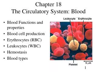
Chapter 18 The Circulatory System: Blood
530 likes | 1.06k Vues
Chapter 18 The Circulatory System: Blood. Blood Functions and properties Blood cell production Erythrocytes (RBC) Leukocytes (WBC) Hemostasis Blood types. Functions and Properties of Blood. Function: Helps cells get nutrition, gets rid of waste Two types of fluid: Blood and Interstitial

Chapter 18 The Circulatory System: Blood
E N D
Presentation Transcript
Chapter 18The Circulatory System: Blood • Blood Functions and properties • Blood cell production • Erythrocytes (RBC) • Leukocytes (WBC) • Hemostasis • Blood types
Functions and Properties of Blood • Function: Helps cells get nutrition, gets rid of waste • Two types of fluid: Blood and Interstitial • Blood is the transported • Interstitial fluid is the diffuser
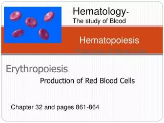
Hematology • The study of blood • (Hemo = blood, -ology = study) • Blood is as unique as your fingerprints • Most cells cannot move around to get oxygen and nutrients and get rid of waste, the blood system does this for them. • INTO the BLOOD- Oxygen from the lungs, nutrients from the small intestines • OUT OF the BLOOD- Waste goes out the kidneys, skin and large intestine
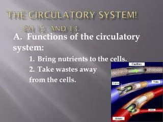
Two Functions of Blood • 1. Transport- oxygen, nutrients, waste • 2. Protection- clotting, immunity = white blood cells (WBC), interferon, complement
Physical Characteristic of Blood • Alkaline: 7.4 pH avg. • 8 % of body weight (if you were a $100 bill, about $8 of you would be blood) • Blood volume: 1.2 gallon female, 1.5 gallon male • Temperature of the blood is a little higher then body temperature, WHY? ______________
Components of Blood • If Blood is spun down (centrifuged), we see two parts: • 1. Plasma- liquid • If this is allowed to clot what remains is called “serum” • 2. Cells- bottom of tube • Cells of the blood are mostly red blood cells (RBCs). The rest, only 1%, is basically platelets and white blood cells (WBCs)
Centrifuging Blood • Centrifuging blood forces cells to separate from plasma • Hematocrit is the % of total volume of cells • Normal crit = females 42 avg., males 47 avg.
Hematocrit Tells Us… • Hematocrit of 40 means that 40 % of the blood is __ __ __s • Does a doctor do a hematocrit to determine the amount of WBCs or platelets? YES NO • Then what is in the buffy coat? ________ • Abnormal: • Low crit =Anemia, decreased ________ • High crit = Polycythemia
Polycythemia • Polycythemia is an excess of RBC • primary polycythemia is due to cancer of erythropoietic cell line in the red bone marrow • Hematocrit of 80% • secondary polycythemia from dehydration, emphysema, high altitude, or physical conditioning • Dangers of polycythemia • increased blood volume, pressure and viscosity can lead to embolism, stroke or heart failure
Plasma Proteins • 3 major categories of plasma proteins • Albumins are most abundant plasma protein • Globulins (antibodies) provide immune system defenses • alpha, beta and gamma globulins • Fibrinogen creates fibrin threads that causes blood to clot
Formation of Blood Cells • Hemopoiesis- Blood making • Lymphoid hemopoiesis occurs in widely distributed lymphoid tissues (thymus, tonsils, lymph nodes, spleen & peyers patches in intestines) • red bone marrow produces RBCs, WBCs and platelets • SEQUENCE: • 1.) Stem cells • 2.) Committed cells • 3.) Precursor Cells • 4.) Final blood cells
Hypoxia: An important concept • Cellular oxygen deficiency is hypoxia (hypo= under, ox= oxygen) • Ischemia is a from of hypoxia due to obstruction of blood flow (as by the narrowing of arteries by spasm or disease) • This can cause cell death (necrosis) • Occurs in many situations: • High altitude • Anemia • Iron or B12 deficiency resulting in anemia • Circulatory problems
Erythrocyte Homeostasis • Classic negative feedback control • drop in RBC count causes hypoxemia to kidneys • stimulation of bone marrow • RBC count in 3-4 days • Stimulus for erythropoiesis • Hypoxia is a stimulus for blood production such as: • low levels of atmospheric O2 (high altitude) • increase in exercise • Circulatory problems • Anemia from Iron or B12 deficiency
Lab Test for RBC creation • Reticulocyte count (“Tic”) measures the rate of erythropoiesis and so can give an indication of RBC creation. • Normal= avg. 1 %--- because about 1% of RBCs are replaced in a day therefore about 1% of the blood is reticulocytes • Low tic count = red bone marrow not working. Cause = nutritional deficiency or leukemia • High tic count = usually good sign, response to iron therapy
Red Blood Cells (RBCs) • RBCs are called erythrocytes (red, cells) • Oxygen-carrying cells • Contains oxygen-carrying protein called hemoglobin • Anatomy- 7.5 microns, biconcave disc- can get into small places • large surface area helps diffusion of gases • No nucleus or organelles so ample space for oxygen • Gets energy anaerobically • So it doesn’t need the oxygen that it’s carrying
Leukocyte Production (Leukopoiesis) • Some lymphocytes leave bone marrow unfinished • go to thymus to complete their development (T cells) • Circulating WBCs do not stay in bloodstream • granulocytes leave in 8 hours • monocytes leave and transform into macrophages • WBCs provide long-term immunity lasting decades
Platelet Production • Cells called megakaryocyte create platelets • Cytoplasm of the cell splits off creating cell fragments that enter the bloodstream as platelets (live for 10 days) • some stored in spleen & released as needed • Platelets are important for _________
Erythrocytes and Hemoglobin Tests • RBC count & hemoglobin concentration indicate the amount of oxygen the blood can carry • hemoglobin concentration of whole blood • men avg. 15; women avg. 14 • RBC count • men 5.5 million avg.; women 5 million avg.
Erythrocyte Death & Disposal • RBCs live for 120 days • membrane fragility -- lysis in narrow channels in the spleen through a process called ____________ • Macrophages in spleen • remove iron from heme • convert heme to bilirubin (yellow pigment) • Why can’t RBCs repair themselves? • Bilirubin is converted to urobilogen in the intestine by bacteria giving fecal matter it’s brown color
Anemia • Causes of anemia • Decreased RBCs or hemoglobin • inadequate dietary vitamin B12 • Or lack of intrinsic factor in the stomach (pernicious anemia) • iron-deficiency anemia • If in males and post-menopausal females check for hemorrhage • aplastic anemia can be drug induced marrow destruction • hemorrhagic anemias from loss of blood • hemolytic anemias from RBC destruction
Anemia • Effects of anemia • FATigue • Heart races • Edema • Low blood pressure • Pallor • Shortness of breath • (Fat HELPS)
Sickle-Cell Disease • Sickle-Cell is hereditary Hemoglobin defect of African Americans • sickle-cell disease individual has shortened life • cell stickiness causes agglutination and blocked vessels • intense pain, kidney and heart failure, paralysis, and stroke • Sickle cell Disease-codominant genetic disorder, resistant to malaria. Why? • Because sickle cells hemoglobin is indigestible to malaria parasites
Blood Types • The surface of RBCs contain genetically determined antigens (antibody generator) these determine the blood types • Major blood types are: ABO and Rh • ABO blood group are based on people that have blood antigens A or B • If you have A antigen on your RBCs you are type A, if you have B antigens you are type B, and type AB has both antigens but if you don’t have any A or B antigens you are type O. • Remember: This applies to incoming blood
Blood Type Diet Theory • Type O is said to be the Original blood type, no antigens. These were the hunters. Diet is meat based. Native Americans are 79% type Os. • Type A is the Agrarian blood type. These are the farmers. Diet is vegetarian.
ABO Blood Typing • Agglutination is the term for blood clumping • For example: type A has agglutination on the left which tells us this blood reacted with anti-A • Type AB has agglutination to both ____ and ______
Blood Swapping • Type AB- universal ABceptor, person can be infused with any type of blood • Type O- universal dOnor, can give their blood to anyone • Caveat: Remember that blood contains other antigens and antibodies than ABO, so specific typing should always be done to avoid Agglutination (clumping) • Agglutination is massive clumping which is distinct from normal clotting (like from a cut)
The Rh Group • Rh or D agglutinogens discovered in Rhesus monkey • If you have the Rh antigen you are Rh+ • If you don’t have the Rh antigen you are Rh- • Anti-D agglutinins are not normally present in blood • formed only in individuals exposed to Rh+ blood • Rh- pregnant woman carrying an Rh+ fetus • no problems result with the first pregnancy • hemolytic disease of the newborn (erythroblastosis fetalis) occurs if mother has formed antibodies & is pregnant with 2nd Rh+ child • RhoGAM is given to pregnant woman to prevent antibody formation and prevent any future problems • Relate disease: Peanut butter should be avoided in ____ peanut butter cup syndrome
Hemolytic Disease of Newborn • Mother’s antibodies attack fetal blood causing toxic brain syndrome from excessive bilirubin in blood from hemolysis • treatment is phototherapy to degrade bilirubin or complete exchange transfusion to replace all the infant’s blood
GRANULAR Neutrophils Eosinophils Basophiles AGRANULAR Lymphocytes Monocytes White Blood Cells (WBCs)WBC are also called Leukocytes (leuko= white, cyte= old cell)-Have nucleus but no hemoglobin_____________________________________
Granular Leukocyte (WBCs) • Granulocytes- when stained these show granules under the microscope • basophils – non-abundant, dark violet granules (<1%) • large U- to S-shaped nucleus hidden by granules • eosinophils - pink-orange granules & bilobed nucleus (2-4%) • neutrophils - multilobed nucleus (60-70%) • fine reddish to violet granules in cytoplasm • Older neutrophils are called polymorphonuclear (PMN) leukocytes or “POLYS” • BEN
Granulocyte Functions • Neutrophils ( in bacterial infections) • phagocytosis of bacteria • releases antimicrobial chemicals • Eosinophils ( in parasitic infections or allergies) • phagocytosis of antigen-antibody complexes, allergens & inflammatory chemicals • release enzymes destroy parasites such as worms • Basophils ( in chicken pox, sinusitis, diabetes) • secrete histamine (vasodilator) • secrete heparin (anticoagulant)
Agranular Leukocyte (WBCs) • These cells do contain granules but do not stainand are small • Monocytes- the cops of the body • The blood is a transport system (cop car) for monocytes as they fight infection in tissues • They go into tissue and become macrophages (macro= big, phage= eater) • Lymphocytes- cytoplasm stains and forms a blue rim around the cell • B cells, T cells and Killer cells
Agranulocyte Functions • Lymphocytes ( in diverse infections & immune responses) • destroy cancer & foreign cells & virally infected cells • coordinate actions of other immune cells • secrete antibodies & provide immune memory • Monocytes ( in viral infections & inflammation) • differentiate into macrophages • phagocytize pathogens and debris
LYMPHOCYTES • Major types are B cells, T cells, and natural killer cells which are major warriors in the immune response • B cells- from Bone marrow • Good at destroying Bacteria • T cells- Formed in bone marrow but matures in the Thymus gland • Good at attacking virus, fungi, cancer cells and Transplanted organs • Killer cells- kill microbes and tumor cells
INCREASED Neutrophils- bacteria Lymphocytes- virus Monocytes-virus, fungus Eosinophils-allergy, parasites Basophils- allergy, cancer DECREASED Neutrophils- radiation, drugs, nutrition deficiency Lymphocytes- chronic illness Monocytes-bone marrow depression Eosinophils-drugs, stress Basophils- pregnancy, stress Increased and Decreased WBCs
WBC Pathology • Leukocytosis- increased WBCs • usually normal • Due to stresses such as microbes and strenuous exercise • Leukopenia- decreased WBCs • Abnormal • due to shock or drug reactions or disease
WBC pathology- Leukemia • Leukemia = cancer of hemopoietic tissue • Uncontrolled WBC production • Subject to opportunistic infection, anemia • Acute Leukemia- = immature WBC • Chronic Leukemia- = too many WBCs • Remission (disappearing) and exacerbation (occurrence) common
Normal and Leukemia Blood Smears • Normal blood ratio = 700 RBCs to 1 WBC
The Action of WBCs • WBCs leave the bloodstream by emigration • Phagocytosis- the process of the WBC eating an invader, a bacteria • Chemotaxis- the process of attracting phagocytes, caused by toxins that are produced by microbes destroying tissue • Lysozymes- enzymes that the phagocytes use to destroy bacteria • A Differential WBC count (Diff) is counting of the different WBC, determining the percentage of each type of WBC helps in the diagnosis
Of Thrombosis and Embolism • Unwanted coagulation • Thrombosis = the formation of a clot of blood within a blood vessel • Thrombosis can create a thrombus • Thrombus = a clot of blood formed within a blood vessel and remaining attached to its place of origin • most likely to occur in leg veins of inactive people • the thrombus may break off causing an embolus • An embolus is an abnormal particle circulating in the blood • An embolus could travel from veins to lungs producing pulmonary embolism • death from hypoxia may occur
Necrosis • Necrosis = Tissue death • Infarct = an area of necrosis in a tissue or organ resulting from obstruction of the local circulation • Infarction is the process of forming an infarct • may occur if an embolus or thrombus blocks blood supply to an organ (MI or stroke) • 650,000 Americans die annually of thromboembolism
GOOD Hemostatis- the process of stopping the bleeding process BAD Thrombosis- blood clotting in vessels forming BLOOD CLOTS Embolism- a blood clot, or air bubble or fat, that blocks a vessel causing Ischemia Hemorrhage- blood loss. Can be from drugs like Celebrex, anticoagulants, or vessel tear COAGULATION(BLOOD COTTING)Blood has the ability to clot together. This is good and bad, depending.
Anticoagulant Drugs • Warfarin or Coumadin (rat poison) may be given to people who have a tendency to produce blood clots, like in atrial fibrillation • Their blood must be monitored for possible hemorrhage • These drugs are antagonists to vitamin K • Streptokinase- produced from streptococcal bacteria is a thrombolytic agent, it dissolves blood clots both good and bad. Used in stroke patients.
Hemophilia • Genetic lack of any clotting factor affecting the patient’s blood coagulation • Sex-linked recessive in males (inherit from mother) • Physical exertion causes bleeding & excruciating pain
Hemostasis - The Control of Bleeding • Platelet plug is formed by pseudopods that adhere to the vessels and contract drawing the vessel together • Fibrin, a sticky protein, comes from the plasma to trap blood cells
Formed Elements of Blood • Never Let Monkeys Eat Bananas- quantities of WBCs