Hematopoiesis
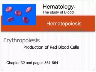
Hematology - The study of Blood. Hematopoiesis. Erythropoiesis. Production of Blood Cells. Production of Red Blood Cells. Chapter 32 and pages 861-864. Lecture outline. I. Functions of blood II. Properties of whole blood III. Whole blood components A. Plasma

Hematopoiesis
E N D
Presentation Transcript
Hematology- The study of Blood Hematopoiesis Erythropoiesis Production of Blood Cells Production of Red Blood Cells Chapter 32 and pages 861-864
Lecture outline I. Functions of blood II. Properties of whole blood III. Whole blood components A. Plasma i. Components of plasma B. Formed elements i. Site of hematopoiesis ii. Hemocytoblast- determined but not differentiated iii. Erythropoiesis a. “Ingredients” b. Process IV. Red blood cell characteristics A. General “need to know” information, indices B. Components within a red blood cell i. Hemoglobin a. Globin chains b. Pyrrole groups c. iron
C. Steady-state maintenance of RBC counts i. Erythropoietin ii. Removal/destruction of RBC a. jaundice • RBC pathologies A. Polycythemia i. Primary ii. secondary B. Anemia i. Defective RBC production a. Iron deficiency b. Aplastic anemia c. megaloblastic ii. Blood loss a. Hemorrhagic (chronic, acute) b. Acquired (transfusion, venom, drugs) c. Inherited (thalassemia, sickle cell, hereditary spherocytosis, G6PD deficiency)
Anemia: a reduced carrying capacity of oxygen • Anemia is not a reduced hematocrit (Hct), because you can have less O2 for other reasons. You can be anemic with normal levels of RBC’s if the hemoglobin is abnormal, and has less iron.
Functions of Blood • Transportation • Gases, nutrients, hormones, wastes, antibodies. • Regulation • Body fluid volume • Body fluid pH • Body T° • Electrolyte levels • Protection from pathogens and bleeding These red blood cells function in oxygen transport Used with permission given by A. Imholtz http://academic.pg.cc.md.us/~aimholtz/
Blood transports O2, CO2, nutrients, wastes, hormones, lipids, etc. The blood also regulates body temperature. Hormones can adjust blood volume, urine output, and maintain pH (which needs to be 7.40). A person with blood pH of 7.35 or 7.45 will not feel well, act strangely. There are buffers in the blood to keep the pH steady, and lungs and kidneys help with this. We also have WBCs in our blood for immune system function, and platelets for clotting.
Blood – Physical Characteristics • Adult ♂ contains 5-6L • Adult ♀ contains 4-5L • T is about 100.4 F • 5 times as viscous as water • What accounts for its viscosity? • pH ranges from 7.35 – 7.45 (slightly alkaline) • What happens at either extreme? • Color ranges—oxygen poor vs. oxygen rich Without O2 With O2 Used with permission given by A. Imholtz http://academic.pg.cc.md.us/~aimholtz/
Adults have 4-6 liters of blood (Let’s call it 5 liters for this class). The heart pumps all of it every 60 seconds. The temperature of our core is higher than the superficial temperature. Blood is 5x thicker than water because of the RBC’s. Increase the numbers of RBC’s, and you will increase viscosity, increase heart workload.
The color of blood is bright red or dark burgundy, depending on oxygenation. Blood is intracellular fluid (RBC’s, 45%) and extra cellular fluid (plasma, 55%). In plasma is mostly water, plus dissolved hormones, proteins, electrolytes. Don’t memorize the next two slides that show everything in the blood; just understand that there are many things in the blood. Water is the solvent. 7g% of the plasma is protein, albumin is the most abundant. Albumin helps keep the water in the plasma by keeping the particle count high. Problems with water leaving the vascular compartment will lead to ascites, so albumin in the blood acts as osmotic support.
Whole Blood Plasma (55%) Formed Elements (45%) • Water (92%) • Plasma Proteins (7%) • Other Solutes (1%) • Red Blood Cells-erythrocytes (99.9%) • Platelets- thrombocytes • White Blood Cells- leukocytes (0.1%) Used with permission given by A. Imholtz http://academic.pg.cc.md.us/~aimholtz/
Transports, organic and inorganic molecules, formed elements, and heat Water (92%) Albumins (60%): Contribute to plasma osmotic pressure; Transport lipids, steroid hormones Plasma Globulins (35%): Transport ions, hormones, lipids; Immune function Plasma Proteins (7g%) Fibrinogen (4%): Essential component of clotting system Other Solutes (1%) Regulatory Proteins (<1%): Enzymes, Hormones Electrolytes: Ions necessary for vital cellular activity. Contribute to osmotic pressure of body fluids. Major electrolytes are Na+ (140meq/L),K+ (3-5 mEq/L), Ca2+, Mg2+, Cl-, HCO3-, HPO42-, SO42- Organic Nutrients: Used for ATP production, cell growth and maintenance; Includes lipids (800mg %) , carbohydrates (80-120mg%) , cholesterol (200mg%) and amino acids Organic Wastes: Carried to sites of breakdown or excretion; Includes urea, uric acid, creatinine, bilirubin, and ammonium ions
Besides albumin, there are other proteins in the blood, such as fibrinogen (inactive form of fibrin). Fibrin is the fiber that grows across a cut and makes a scab. Other proteins include regulatory and proteolytic enzymes. Other substances in blood include glucose, lipids, cholesterol, bilirubin (waste product of RBC destruction), and creatinine (waste product of kidney). We check those levels in the blood to ascertain kidney function. There are lots of solutes in the blood!
BLOOD ANALYSIS TERMINOLOGY • You need to measure hematocrit (Hct), hemoglobin (Hgb), and do a RBC count. With this information, you can then calculate MCV, MCH, and MCHC. By the way, RBC = corpuscle. • MCV(mean corpuscular volume) calculated as MCV = Hct / RBC count. Normal is 80-100 µm • MCH (mean corpuscular hemoglobin) calculated as MCH = Hb / RBC count. Normal is 32 pcicograms (10 -12 micrograms). There is not much, but hemoglobin is still the most abundant protein in RBC’s. • MCHC (mean corpuscular hemoglobin concentration) is the ratio of MCH to MCV. It is calculated as MCHC = Hb/Hct. Normal is 34% • MCD (mean corpuscular diameter) normal is 7-8 µm wide, 2 µm thick.
100 ml Red blood cell- need to know information • Hematocrit: % volume of blood that is red cells • Men ~45% (42-52%) • Women ~40% (37-47%) • Hemoglobin concentration -Wt of Hb in 100 ml blood • 15-16 (male) gm Hb/100 ml blood • 13-14 (female) gm Hb/100 ml blood • Oxygen carrying capacity: • gm Hb/100 ml blood * 1.34 ml O2/gm Hb • ~21 ml O2/ 100 ml blood for men • ~19 ml O2/ 100 ml blood for women
Red Blood Cell “Indices” • Mean Corpuscular Volume • MCV 80-100 m3 • Mean Corpuscular Hemoglobin • 32g (10-12; picogram) • Wt of Hb in a single RBC • Mean Corpuscular Hemoglobin Concentration • 34% • Concentration of Hb to volume in a single RBC (i.e., solute/solvent). • Mean Corpuscular Diameter • MCD 7-8 m • Color Index • CI 0.9 – 1.1 A “corpuscle”
COLOR (Chromasia; indicates how much Hgb is there) Normal is 1.0 • Normochromic is 1 • Hypochromic is less than 1. Can’t have hyperchromic RBC’s. A disorder that appears hyperchromic is • Hereditary Spherocytosis. It is the most common inherited disorder that affects the RBC membrane. RBC comes out of marrow normal, but with time, the membrane is lost, but none of the contents are lost, RBC gets smaller and concentrated, looks redder. Thus, it could look hyperchromatic.
SIZE • Normocytic is normal size. • Microcytic (small) • Macrocytic or Megaloblastic (big) • SHAPE • Poikilocytosis is abnormal shapes, such as sickled or sphaerocytes. Cells with abnormal size or shape will be removed from circulation faster, get anemia.
Terminology • Chromasia- indication of MCH • Hyper/hypochromic • normochromic • Anisocytosis – cells of abnormal size; indication of MCV • Microcytic • Macrocytic/ Megalocytic • normocytic • Poikilocytosis – cells of abnormal shape • Spherocytosis • Sickle cell • Echinocyte
CELL COUNTS • We have 5 liters of blood, 25 trillion RBC’s, 4.5 million in one microliter. We need the rate of old blood cells that die to match the rate of new ones being made. RBC’s are made by stem cells in bone marrow. Too many RBC’s is polycythemia, too few is anemia. RBC shape should be biconcave because that increases surface area for O2 binding and allows it to be flexible to get through capillaries. When a RBC gets older, its membrane is less flexible, gets stuck in liver and is detected and destroyed. Because of the shape increasing surface area, there is rapid gas exchange across cell membranes for wastes and O2. You know that hemoglobin binds O2, but CO2 can also bind to hemoglobin, but only if O2 is not there.
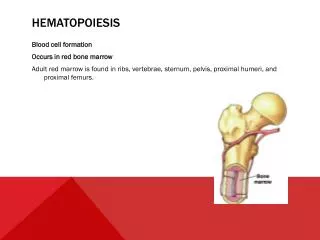
SITES OF HEMOPOIETIC ACTIVITY • An embryo (< 3 months) makes blood in yolk sac • Fetus (> 3 months) makes blood in the liver • At birth, makes blood in the bone marrow • Blood that is made in the bone marrow is specifically made in the axial and appendicular skeleton until the age of 20. Thereafter, it can only be made in the axial skeleton, mainly in the sternum and iliac crest.
Bone marrow Yolk sac Vertebra Liver Sternum Rib Spleen Femur Tibia 20 3 1 ADULT FETAL MONTHS Let’s talk about formed elements-Sites Of Hemopoietic Activity Axial skeleton Appendicular skeleton
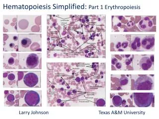
ERYTHROPOIESIS (MAKING A RBC) • A stem cell that is completely undifferentiated yet is called a pleuripotent stem cell. • A pleuripotent stemcell can differentiate into any cell type: nerve, muscle, blood, etc. Since we are discussing blood cell formation, we will focus on a pleuripotent stem cell that differentiates into a Hemocytoblast.
Hematopoiesis • The “pluripotent hemopoietic stem cell” or “hemocytoblast” is the precursor of all blood cells. • Found in bone marrow. • Undergoes mitotic divisions • daughter cells differentiate • This is the source of all new blood cells. • The “colony-forming unit, myeloid stem cell line” • The “colony forming unit, lymphoid stem cell line” Myeloid stem cell
Hemocytoblasts can differentiate into any blood cell type. Therefore, they are not pleuripotent anymore, but they are still multipotent (they are “determined”, but not completely differentiated). A hemocytoblast will continue to differentiate into one of two cell types: • Lymphoid line generates B and T cells • Myeloid line generates red and all other blood cells. Scientists doing stem cell research have found a way to turn a multipotent cell back into a pleuripotent cell by just Inserting four genes into a multipotent cell!
To make a good RBC, you need to start with good ingredients: a good hemocytoblast, proper nucleotides, folic acid, vitamin B12, and other vitamins. You also need growth inducers, differentiation markers (signals), amino acids (Adenine, Thymine, cytosine, guanine), heme (a pyrrole ring and globin proteins), and iron.
The Recipe for Normal Erythropoiesis • Healthy stem cells • Growth inducers (interleukins) • Differentiation inducers • Cell Division (Mitosis) • DNA replication- vitamin B12 and folic acid coenzymes • building blocks of DNA • (Adenine, Thymine, Cytosine, Guanine) • Hemoglobin Synthesis • amino acids, heme and iron
Newly formed red blood cells have to get from the bone marrow into a blood vessel. To do this, they squeeze through the endothelial cells of the vessel, but their nucleus cannot fit, so it gets pinched off. The new RBC in the bloodstream has a little bit of endoplasmic reticulum and bits of DNA deposits left over from where the nucleus was pinched off, so a brand new (immature) RBC in the bloodstream is called a reticulocyte. Thus, RBC’s are in an immature state when they are released into the bloodstream. It takes about 1-2 days for these endoplasmic bits to dissolve. Until then, you can see the difference between a reticulocyte and a mature RBC under the microscope when looking at a blood smear. Reticulocytes are immature red blood cells. Only about 1% of the RBC’s should be reticulocytes. If there are more, it indicates a problem.
Hemocytoblast Erythropoiesis-5-7 days Pluripotent stem cell Myeloid Stem Cell Could become an RBC or several types of WBC Destined to become an RBC; “more” determined but still not differentiated Various stages. Actively synthesize Hb Just lost its nucleus. Enters the circulation – should be no more than 1% among all RBCs. Mature RBC--Differentiated. (After a reticulocyte has been in the blood stream for 1-2 days) What is a functional advantage of the fact that the RBC lacks mitochondria?
As long as a RBC is flexible, it can weave around the web of reticular fibers in the liver and spleen without getting stuck. Those that get stuck are phagocytized by macrophages. RBC’s that are old or hemoglobin abnormalities (sickle cells) tend to be less flexible, and are caught in reticular fibers and destroyed. This can also happen to reticulocytes. • This process takes 7 days: hemocytoblast myeloid stem cell reticulocyte RBC • Up until the reticulocyte is released, it retains its shredded endoplasmic reticulum, trying to undergo protein synthesis to make hemoglobin as much as possible.
Rate of Production Bone Marrow Steady State Rate of Removal Liver, Spleen Red Blood Cell physical properties and components • Most abundant blood cells – ¼ of body cells • 4.5 - 5.5 x 10 6 cells / mm3 • Why a biconcave disc? • Provides a large surface area for O2 entry/exit • Enables them to bend and flex when entering small capillaries • simply membranous bags of hemoglobin, a protein found in extreme abundance in RBCs, which binds and transports O2 and CO2
Erythropoiesis is stimulated by a hormone called EPO, which is secreted by the kidney. • In conditions where there are not enough RBC’s in the body (e.g. Erythroblastosis Fetalis), oxygen levels will decrease. The kidneys will sense that, and produce EPO, which stimulates stem cells to divide faster, and hastens the release of immature RBCs into the bloodstream, so we will see more reticulocytes in a blood smear.
When the RBC is fully mature, it lacks organelles. This is good, because it can transport more O2 if it does not contain any mitochondria, which consume oxygen. However, there is a disadvantage, to not having a nucleus and other organelles: a RBC can’t repair any damage or express new proteins. That’s why they only live 120 days; they accumulate damage, lose their flexibility, and get destroyed. A true RBC count should be at a steady state, new ones replace dying ones.
I want my oxygen! O2 O2
A Hemoglobin molecule consists of three parts • Globin chains (proteins from gene expression) • Heme Group • Pyrrole ring Iron Heme Group
Hemoglobin • Large protein consisting of 4 polypeptides • 2 chains and 2 β chains • Each chain contains a single molecule of heme, an iron-containing pigment • The iron ion in heme is able to reversibly bind an oxygen molecule thanks to the surrounding globin chains. • Meaning, O2 can bind to Hb at the lungs and then be released at the tissues • Note the 2 chains and 2 β chains. Notice how each has an associated heme molecule with an iron atom. • Based on the above, how many molecules of O2 can each Hb protein bind?
GLOBIN CHAINS • To make the globin chains, we need genes. If there is a defect in the gene, the globin chains are defective, as in the case of sickle cell disease. Since it is the iron that binds the oxygen, why do we need globin at all? Because iron binds to oxygen so strongly, it will never let go unless hemoglobin is there to move its structure to block the magnetism of the iron. We need for iron to bind strongly to the oxygen in the lungs. When there is no oxygen on a hemoglobin molecule, the globin chains move a little, exposing the iron so it can grab some oxygen while in the lungs. Once the iron is bound to oxygen, the globin chains move a little, decreasing the hold of iron onto the oxygen, so that the first oxygen-depleted cell that it comes close to can pull the oxygen molecule off of the hemoglobin complex. This is considered reversible binding of oxygen; hemoglobin has an affinity for oxygen in lungs, and low affinity to oxygen in the tissues.
There are different types of globin chains. In an adult, there are 2 alpha globin chains and 2 beta chains. Therefore, adult hemoglobin is called A2B2. Since each globin chain is a protein, and there are four proteins bound to each other, hemoglobin has quaternary structure. An embryo has embryonic hemoglobin, called A2E2. An embryo does not have working blood vessels yet, since oxygen is coming in from the placenta. Therefore, an embryo needs Hgb with a higher affinity for oxygen than mom’s A2B2, to rip the oxygen off mom’s hemoglobin. When the embryo develops into a fetus, its blood vessels get bigger, have closer proximity to mom’s blood vessels, needs a little less affinity than embryonic hemoglobin, but the fetus still needs to have hemoglobin that has a higher affinity for oxygen than A2B2, so fetal hemoglobin is A2G2. Around the time of birth, the baby’s hemoglobin becomes A2B2. Once a baby is breathing on its own, it needs hemoglobin with lower affinity. Therefore, the order of affinity for oxygen of the different types of hemoglobin is HgbE (A2E2), HgbF (A2G2), then HgbA (A2B2).
Globin Chains • Why not just use iron/heme group? • Determine Hb’s affinity for oxygen • Are expression products of different genes (chromosomes 11, 16) • Different genes are expressed throughout life and have different affinities for oxygen • Embryonic Hb (HbE) 22 (before 3 months gestation) • Fetal (HbF) 22 (replaced within 6 months of birth) • Adult (HbA) 22 = heme group (+ iron) = alpha chain = Beta chain
HEME GROUP • Within each globin chain is a heme group. A heme group consists of a pyrrole ring, with an iron atom in the middle. Since there are four globin chains per hemoglobin molecule, each Hgb has 4 irons.
MAKING HEMOGLOBIN • Hemoglobin is made in the mitochondria of the erythrocyte while it is developing (in the proerythroblast stage). Once iron is added to the pyrrole ring, the entire structure is called heme. When you add the four globin chains to heme, it is now called hemoglobin. When a macrophage phagocytizes a RBC, the hemoglobin is taken apart into its components. The iron and globin are recycled, but the pyrrole group is cannot be reused, so it needs to be eliminated from the body as a waste product.
The making of a pyrrole ring in proerythroblast mitochondria Alpha and Beta Globin polypeptide chains are synthesized at the ribosome in the cytoplasm. Completed porphyrin rings are sent to meet them in the cytoplasm, where they all bind to form Hb.
We get iron from our diet. It is absorbed from the intestine and released into the plasma, where it binds to a plasma protein called apotransferrin (when it is not bound to iron) or transferrin (when it is bound to iron). Since we have said that the plasma protein has bound to the iron, we will now call it transferrin. The transferrin protein takes the iron to cells in the body that need iron, or the iron is stored intracellularly in two different forms. • Ferritin is a protein within a cell that has bound onto the iron. This same protein, when unbound, is called apoferritin. • Hemosiderin is a complex in cells that binds to iron and does not release it for use very easily; it is very insoluble. Macrophages that phagocytize RBC’s tend to accumulate hemosiderin deposits. Too much hemosiderin in a cell or in tissues is toxic.
Iron • Obtained in the diet • Released into plasma • Binds to protein called “apotransferrin” • Travels in circulation as transferrin • Delivered to cells needing iron or stored intracellularly in two different forms • Apoferritin- to form iron- bound ferritin • Hemosiderin-extremely insoluble; toxic to cells; iron over-load • Ferrous (reduced; +2) form binds indirectly to oxygen • Ferric (oxidized; +3) form cannot bind oxygen • Hb with iron in ferric form is called methemoglobin
IRON HAS SEVERAL DIFFERENT STATES • Ferrous (reduced +2) form binds indirectly to oxygen. We need it in this state. • Ferric (oxidized +3) form cannot bind to oxygen. Hgb with iron in ferric form is called methemoglobin. • We have proteins to convert iron from its ferric to the ferrous state. There are some household products and pesticides that change ferrous to ferric, and our body may not enough available energy to convert it back to the ferrous form we can use.
ERYTHROPOIETIN (EPO) • EPO (erythropoietin) is a hormone; 90% of EPO is made in the kidney, 10% is made in the liver. It stimulates all the stem cells of blood, many of which develop into RBCs. The RBCs are ready to exit the bone marrow and enter the circulation in 5 days, plus another 2 days of maturation within the circulatory system. If you lose kidney function, you can become anemic. EPO is released by the kidney in response to low oxygen levels in the tissues (hypoxia).
Steady state RBC count-Erythropoietin (EPO) • What is it? A hormone • What is its source? 90% from kidney, 10% from liver • Conditions for its secretion? Kidney senses hypoxemia (low oxygen; is most essential regulatory of RBC production) • Erythropoietin is always present in the plasma we would be anemic without it! • What are its actions? • Promotes release of reticulocytes • Stimulates stem cell mitosis • Increase in red cell number in 5 days • Synthetic EPO /Recombinant Human EPO—”Ecrit” “Eprex” “Dynepo”
Chemotherapy for cancer patients targets rapidly dividing cells, especially the hair (causes hair loss), stomach lining (causes nausea), and the bone marrow (causing low RBC count, and fatigue). They are given artificial EPO to offset the anemia. Athletes might take EPO (illegally) for “blood doping”. It causes an increase in RBC production, leads to more O2, more ATP, more energy, but it thickens blood, can cause heart attack. Sports authorities have been using a drug test on athletes who use a form of EPO made from bacteria; the bacterial particles are detectable on blood tests. Now, these unscrupulous athletes are getting clever: someone has manufactured human EPO that cannot be detected in these drug tests. This human EPO is approved for medicinal use in Europe, and it is in America on the Black Market. So now, sports authorities do a RBC count. If the RBCs are present in excess of set limit before the race starts, they are disqualified. Some of these athletes get away with it by training with human EPO and donating blood before the race.
If you want to climb a high mountain, you can’t just climb to the top in one day. There is less oxygen pressure at high altitudes, so RBCs can’t bind oxygen as well. What you do is go to a base camp, part way up the mountain, and stay there for 2 weeks, so the kidney can release EPO to stimulate RBC production. Then you go up the mountain to the next base camp for 2 weeks. Then you can go to the top, when the new cells can bind to oxygen better.
RBCRemoval • Lifespan approx. 120 days • RBCs are subjected to incredible mechanical stress. • Narrow capillaries • Limited ATP stores for replacement of worn parts • Why are they unable to synthesize replacements for damaged parts? • Membrane fragility • Macrophages in liver and spleen remove old RBCs • Contents destroyed or recycled Above, we have a macrophage phagocytizing multiple RBCs • How many RBCs did it engulf?
Three different cells participate in destruction of RBCs • RBC • Macrophage • Hepatocyte