Download
1 / 43
500 likes | 1.09k Vues
What is? Hematopoiesis. Bone Formation. Fetal INTRAMEMBRANOUS OSSIFICATION ENDOCHONDRAL OSSIFICATION From birth to young adult APPOSITIONAL (flat, short, irregular bones) Increase diameter for ALL bones INTERSTITIAL (EPIPHYSEAL PLATE) Increase length. SKELETAL TISSUES CHAPTER 7 .

E N D
Bone Formation • Fetal • INTRAMEMBRANOUS OSSIFICATION • ENDOCHONDRAL OSSIFICATION • From birth to young adult • APPOSITIONAL (flat, short, irregular bones) • Increase diameter for ALL bones • INTERSTITIAL (EPIPHYSEAL PLATE) • Increase length
SKELETAL TISSUESCHAPTER 7 By John McGill Supplement Outlines: Beth Wyatt Original PowerPoint: Jack Bagwell
MACROSCOPIC STRUCTURE: Long Bones • DIAPHYSIS • Shaft • Composed of Compact Bone • EPIPHYSES • Both Ends Composed of Cancellous Bone • ARTICULAR CARTILAGE • “Joining Cartilage” • Covers Epiphyses (Thin Layer) • Provides Cushioning at Joints
MICROSCOPIC STRUCTURE OF BONE: COMPACT BONE: • HAVERSIAN SYSTEMS (OSTEONS) • Microscopically, Compact Bone is Composed of Haversian Systems • Haversian Systems: Microscopic Structural Units of Compact Bone
BONE (MICROSCOPIC VIEW) osteocyte in lacunae canaliculi Haversian canal ossified matrix (lamellae)
CANCELLOUS BONE: TRABECULAE • Trabeculae: Needlelike Pieces of Bone (Surround Spaces) • Contains Osteocytes • How Cancellous Bone Gets Its Blood Supply: • Periosteum Bone Marrow Openings in Trabeculae
Types of BONE CELLS • OSTEOBLASTS • Bone-Forming Cells • Location: Periosteum (Primarily) • OSTEOCLASTS • Bone-Destroying Cells • Location: Endosteum (Primarily) • OSTEOCYTES • Bone Cells (Mature Osteoblasts) • Locations: • 1) Compact Bone: Lacunae • 2) Cancellous Bone: Trabeculae
BONE MARROW is MYELOID TISSUE Myeloid tissue is a biologic tissue with the ability to perform hematopoiesis.
BONE MARROW TYPES: RED MARROW • DESCRIPTION/FUNCTIONS • Red in Color Because Functions in Hematopoiesis • LOCATIONS • Children: All Bones Contain Red Marrow • Adults: Certain Bones Contain Red Marrow • Flat Bones of the Skull • Sternum, Ribs, Vertebrae • Pelvic Bones • Epiphyses of Humerus and Femur
BONE MARROW TYPES: YELLOW MARROW • DESCRIPTION/FUNCTIONS • Yellow in Color Because Contains Largely Adipose Tissue • Yellow Marrow Was Once Red Marrow, Now Yellow B/C • It No Longer Functions in Hematopoiesis • LOCATIONS • Most Bones in Adults Contain Yellow Marrow
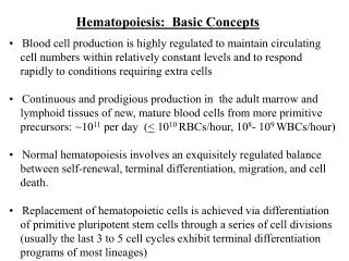
Functions of Bones • Support • support the weight of the rest of the body • Protection • protect the delicate body parts • Movement • muscles attach to bone and allow movement • Mineral storage • calcium, phosphorous, and other minerals are stored in the bone • Hematopoiesis • red marrow plays an important role in the formation of red blood cells, some flat bones also play a role here
Fetal Bone FormationOsteogenesis • The cartilaginous skeleton is changed to bone in one of two ways: • Intramembranous ossification • Endochondral ossification
#1. INTRAMEMBRANOUS OSSIFICATION • How Bones Form in the Fetus • DEFINITION • “Within Membrane Bone Formation” • Method by Which Flat Bones Form • MECHANISM • Connective Tissue Membrane • Cells Develop Into Osteoblasts • Secrete Organic Matrix and Collagenous Fibers • Calcification Occurs • Intramembranous bone formation in a fetal pig skull. • Flat bones of the skull develop by IO. • Embryonic mesenchyme cells form a membrane (Mes) & • differentiate into osteoblasts that • form bony spicules or cancellous bone (CsB). • Eventually osteonsform.
ENDOCHONDRAL OSSIFICATION DEFINITION “Within Cartilage Bone Formation” Method by Which Most Bones Form MECHANISM Cartilage Model Periosteum Forms Cells Develop Into Osteoblasts Secrete Organic Matrix and Collagenous Fibers Calcification Occurs *Note: In Both Types of Ossification: OsteoclastsResorb Bone Forms Medullary Cavity, Spaces in Cancellous Bone Embryonalhyaline cartilage is model for bone formation. Osteoblasts begin to calcify the cartilage Osteoblasts and osteoclasts are constantly reshaping the bone Epiphyseal plate is site of continued bone growth; indicates the bone is not yet mature. #2. ENDOCHONDRAL OSSIFICATION
Bone Growth - Animation • http://www.anatomy.gla.ac.uk/fab/tutorial/generic/bonet.html
BONE GROWTH AND RESORPTION • How Bones Increase in Size after Birth • FLAT BONES (Also Short, Irregular Bones) grow by APPOSITIONAL GROWTH • Growth By Adding to the Surfaces • LONG BONES grow in length by EPIPHYSEAL PLATE • EpiphysealPlate: Layer of Hyaline Cartilage That Lies B/T Epiphyses and Diaphysis • Didn’t Ossify During the Fetal Period (Purpose: To Allow Bone Growth in Length) • Epiphyseal Plate 1) Thickens and 2) Ossifies Repeatedly • When Growth in Length is Complete, Cells in EP Stop Mitosis and the Entire Plate Ossifies, What Remains is Epiphyseal Line
EpiphysealPlate The epiphyseal plate allows for growth in bones.
GROWTH IN DIAMETER – COMBINED ACTION OF OSTEOBLASTS AND OSTEOCLASTS • Osteoblasts (Periosteum) Build New Bone on the Outer Surface • Osteoclasts (Endosteum) Destroy Bone from the Inner Surface of the Medullary Cavity (Enlarges Med. Cavity) • http://www.youtube.com/watch?v=6Cn4uusbGk8
BONE GROWTH AND RESORPTION • BONE GROWTH AND RESORPTION THROUGHOUT LIFE • Both Growth and Resorption Go On Throughout Life, But at Different Rates • From Infancy Young Adulthood: Growth EXCEEDS Resorption (Bones Grow and are Thick) • During Late 20’s/Early 30’s: Growth EQUALS Resorption (Bones Remain Relatively Constant) • From Mid 30’s/Early 40’s Old Age: Resorption EXCEEDS Growth (Bones Become Thinner, More Susceptible to Fracture and Disease)
BONE GROWTH AND RESORPTION • BONES RESPONSE TO STRESS • Bone Stress = Weight Bearing Applied to Bones • Bone Stress Increases the Activity of the Osteoblasts (Helps Offset the Effects of Aging on Bones)
REPAIR OF BONE FRACTURES • FRACTURE: A Break in the Continuity of Bone
FRACTURE HEALING • VASCULAR DAMAGE • Damage to Blood Vessels
FRACTURE HEALING • FORMATION OF FRACTURE HEMATOMA • Blood Clot Forms in the Area of the Fracture in Order to Stop Bleeding
FRACTURE HEALING • FORMATION OF CALLUS TISSUE • Thickened Repair Tissue That Binds the Ends of the Bones Together (Reason That the Fracture is Aligned and Immobilized)
FRACTURE HEALING • REPLACEMENT BY BONE • Callus Tissue Becomes Bone (Action of Osteoblasts), Remodeled by Osteoclasts
CARTILAGE • CHARACTERISTICS • MATRIX • FIRM/FLEXIBLE GEL • PROTEIN FIBERS • COLLAGENOUS • CELLS • CHONDROCYTES • Chondrocytes Lie in Lacunae • AVASCULAR: Oxygen and Nutrients by Diffusion
Similarities and Differences Bone Cartilage
Similarities and Differences Collagen Ground substance/matrix Cells Protection Connective tissue Mitosis (both grow) Appositional, insterstitial Lacunae Bone Cartilage
CARTILAGE: Types • Hyaline • Elastic • Fibrocartilage
HYALINE CARTILAGE • Most Abundant and Common • Shiny • Semitransparent • Locations: • Articular Cartilage • Costal Cartilages • Cartilage Rings in Trachea and Bronchi • Tip of Nose
ELASTIC CARTILAGE • Has Fewer Collagenous Fibers Compared to Hyaline • In Addition, Contains Elastic Fibers • Locations: • External Ear • Epiglottis • Eustachian Tube
FIBROCARTILAGE • Cartilage With the Most Collagenous Fibers • Locations: • Symphysis Pubis • Intervertebral Disks • Menisci in Knee
GROWTH OF CARTILAGE • INTERSTITIAL (ENDOGENOUS) GROWTH • DEFINITION: “Growth From Within” • OCCURS WHEN: During Childhood and Adolescence • APPOSITIONAL (EXOGENOUS) GROWTH • DEFINITION: “Growth by Adding to the Surfaces” • OCCURS WHEN: During Adulthood