Download
1 / 57

E N D
HematopoiesisFrom Greek haima for blood and poiein, to make.Dynamic and Complex Developmental Process of the Formation of New Blood Cells.Early Intra-Embryonic Site: Paraaortic Splanchnopleura (PAS) and AGMHematopoietic Stem Cells (HSC) arise in Blood Islands of the Yolk Sac, PAS and AGM, Enter Embryonic Circ and Colonize Newly Formed Liver Rudiment.Primitive (prior to the dev of Fetal Liver) vsDefinitive (after). Bone Marrow for post-natal. .
Properties of HSCSelf-Renewal Potential: Progeny Identical in Appearance & Differentiation PotentialMultilineage Differentiation Capacity: Lymphopoietic & Myelopoietic PotentialMarrow Repopulating Ability: Colonize BM of lethally irrad an & reconstitute entire hematopoietic system
Murine Hematopoiesis Lymphohematopoietic progenitors simultaneously identified in PAS and YS at 8.5 dpc. CFU-S appear in YS and AGM at late 9 dpc (AGM >YS). LTR-HSC first in AGM at 10 dpc, before YS & FL: controversal By 12 dpc, the liver is the predominant site IMPORTANCE OF THE MICROENVIRONMENT
HUMAN FETAL HEMATOPOIESIS At 2-8 wks, primitive nucleated erythroid cells in YS, contain Hb, not fully developed. During the 2nd mth, extramedullar hematopoiesis: YS cells migrate to liver. Spleen contributes to hematopoiesis. During the 4th mth, medullary hematopoiesis develops to take over during the 5th month.
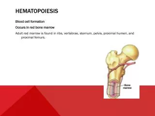
HUMAN HEMATOPOIESIS after BIRTH At birth, liver and spleen cease production of blood cells. Hematopoiesis occurs in the red BM of bones. At 4 yrs of age, hemato activity begins to move to the axial skeleton (flat bones, skull, ribs, sternum, clavicle, vertebrae, pelvic bones) and proximal ends of long bones (humerus and femur). Move complete by age 18. Remaining marrow cavities are replaced with fat (yellow BM).
HUMAN ADULT HEMATOPOIESIS By age 40, the marrow in sternum, ribs… is composed of equal amounts of hematopoietic tissue and fat. Extramedullary hematopoiesis may occur: if the BM no longer functional, induced hematopoiesis (enlarged liver and spleen). Stem Cells make up 10% of cord blood cells and <1% of all adult blood cells
HSC assays in vivo and in vitro Long-term reconstitution of irradiated mice (ablated or severely impaired endogenous hematopoiesis) Cell surface markers (mAbs + Flow Cytometry & Cell Sorting) Spleen Colonies formed 12 days after injection of sorted HSC into a lethal irrad host in theColony-Forming-Units- Spleen (CFU-S) Assay. Not Possible in Humans! Mouse-Human Chimaeric Models (Hu FL, Bone, Thymus or PB transplanted in SCID mouse) Long-Term Culture-Initiating Cell (LTC-IC) Assay: HSC containing cell populations plated on preformed layer of stroma cells. Cultures scored for colony-forming cells. Cobble-stone-Area-Forming Cell Assay: Test population plated on preformed stromal cell monolayer. Cultures scored = f(time) for presence of cobble-stone areas (35 days = HSC)
Neutrophils 60-80% in Peripheral Blood Differentiation, proliferation and maturation takes place in the red BM. Developmental stages: Myeloblast (prolif), Promyelocyte (big,prolif), Myelocyte (prolif),Metamyelocyte (matur), band (matur), segmentedNeutrophil(matur) Cell and Nucleus Sizes with maturation, Nuclear Indentation Primary (nonspecific) granules: promyelocyte Secondary (specific) granules: myelocyte
Neutrophils: Functional Development Attracted to the site of inflammation by Chemotaxis to exert Phagocytosis Primary granules contain Myeloperoxidase required for intracellular microbial killing Secondary granules contain Alkaline Phosphatase and other compounds necessary to kill and degrade toxic, infectious or non-self agents
Neutrophils Steps ofPhagocytosis Opsonization: Greek = to prepare for dining Coating with Ig and Complement for ingestion. PMN mb pseudopods envelop the microbe, forming a vacuole called a phagosome within the cytoplasm. Fusion phagosome/lysosomes Lytic enzymes kill & digest the foreign agent
Monocytes/Macrophages Circulating monocytes (3-7% of WBCs) become macrophages (larger) when migrate into tissue, display eccentrically placed and round nucleus Maturational Changes: Monoblast (id myeloblast) Promonocyte, N:C ratio w/ maturation, lobular and irregular nuclear shape, nucleoli disappear, coarse chromatin pattern Monocyte cytoplasm++ w/ vacuoles
Monocytes/Macrophages: Function Numerous MF in “filter”organs (spleen, liver, lungs, lymph nodes = Mononuclear Phagocyte System) = Body Defense composed of phagocytic cells, Cytokine Producers Special Names: MF in liver, Kupffer cells, osteoclasts in bone, microglial cells in nervous tisssue, Langerhans’ cells in epidermis (APC), glomerular mesangial cells in kidney, pulmonary alveolar MF in lungs.
Stem Cell Markers IL-3R (CD123) Hemopoietin Receptor Superfamily c-KIT (CD117)RTK, SCF is ligand Sca-1 (Mouse) Stem Cell Antigen Flk-2/Flt3 receptor RTK on ST-HSC (transient multilin reconstituting potential) CD-34 (Human) Sialomucin (CD45-RA, PSGL-1 or CD162, PCLP-1) ADHESION RECEPTORS:b1-integrins (ligand = Vascular Cell Adhesion Molecule (VCAM-1 or CD106) b2-integrins (ligand = Inter cellular Adhesion Molecules or ICAMs) (cell/ECM) CHEMOKINE RECEPTORS
KIT and SCF c-KIT gene id as human counterpart of the HZ4 feline sarcoma virus harboring the v-KIT gene, related to PDGFR and CSF-1R. Cloning of loci dominant White spotting (W) and Steel (Sl) present in natural mouse mutants lead to the id of c-KITR and c-KIT ligand or Stem Cell Factor (SCF). Isoforms of KIT (4aa upstream of TM domain): ligand-indepen dent constitutive phosphorylation observed in isoform -4aa. SCF: Stromal Cells, Fibroblasts, Endothelial Cells. Bivalent Dimer, Membrane-associated or soluble form.
CYTOKINE RECEPTOR FAMILY Classification based on structural motifs in EC domains, from Rane & Reddy, Oncogene (2002) 21:3334 1. The gp130 Family:IL-6, IL-11, Ciliary Neurotrophic Factor (CNTF), oncostatin M, Leukemia Inhibitory Factor (LIF) all signal through a common b chain called gp130, but cytokine-specific ligand binding a. Conserved EC domain (200 aa), 4 conserved Cys in the N-terminal region and a trp-Ser-X-Trp-Ser (WSXWS) motif proximal to the TM domain. 2. The IL-2 Receptor Family: IL-2R 3 sub-units, a, b and g. Gamma chain shared by IL-4R, IL-7R, IL-9R and IL-15R. 3. The gp140 Family: IL-3R, IL-5R, GM-CSFR with unique a chains and shared b chain. Gp140 is the signal transducing unit. 4. The Interferon (IFN) Family: IFNa R, IFNg R, IL-10 R. 5. The Growth Hormone (GH) Family: GH R, Prolactin R, EPO R, TPO R.