Download
1 / 22
230 likes | 315 Vues
Explore the intricate processes of hematopoiesis, the formation of blood cells in the red bone marrow, and hemostasis, the mechanism of blood clotting. Learn about RBC development, Erythropoietin function, and the clotting cascade in response to vessel injuries.

E N D
(1) Hematopoiesis • Blood Cell Formation • Occurs in Red Bone Marrow • Cell Development: Hemocytoblast (original stem cell) Lymphoid Stem Cell Myeloid Stem Cell • Erythrocytes • Basophils • Eosinophils • Neutrophils • Monocytes • Platelets • Lymphocytes
(2) Red Blood Cell Development • RBC’s have no nucleus… • Synthesize hemoglobin during development • Increase in hemoglobin Eject Organelles • Results in Biconcave Shape • Erythropoeitin:Protein which controls rate of RBC production. • Circulates in blood • Targets bone marrow • Produced in liver + kidney • Decrease in Oxygen Increase in Erythropoeitin
(3) Rate of RBC Production (Erythropoeitin Release More RBC’s) • Stimulus: • Change in RBC count • Change in Oxygen availability • Change in demand for Oxygen • Scenario: Decreased RBC count… • Reduced Oxygen in blood • Kidney releases erythropoeitin • Red bone marrow stimulated • RBC’s produced
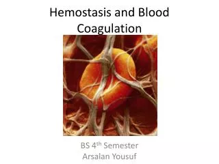
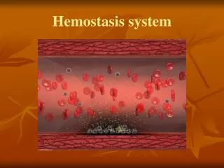
(4) Hemostasis (Blood Clotting) • Stimulus: Injury to blood vessel. • Events: 1. Collagen fibers exposed, Platelets adhere. 2. Platelets release serotonin to constrict vessels, More platelets seek wound. 3. Thromboplastin released by damaged vessel cells. 4. PF3 (Phospholipid) binds with Thromboplastin + Calcium BEGIN CLOTTING CASCADE!
Clotting Cascade (Prothrombin Activator): • Prothrombin converted to Thrombin. • Thrombin joins Fibrinogen proteins to form larger polymer (Fibrin). • Fibrin forms a mesh-like trap to catch RBC’s. • Vessels Constrict and ruptured edges are closed together.
Initial Injury Clotting Cascade Begins Prothrombin changed to Thrombin by Prothrombin activator Thrombin binds with Fibrinogen to form Fibrin… Finally the mesh-like net is formed to catch blood cells
(5) Thrombus vs. Embolus • Thrombus Clot in wall of blood vessel • Embolus Free Floating Thrombus • Causes: • Rough blood vessel tissue from burns, ruptures, fat. • Slow blood flow (low levels of activity) • Aspirin: Common over the counter anticoagulant.
(6) Thrombocytopenia (Hemostatic Disorder) • Low platelet count & Slow Clotting Rate • Typical of bone marrow cancer and/or radiation patients. • Caused by damaged liver Cannot make clotting factors.
(7) Hemophilia (Hemostatic Disorder) • Low Clotting Factor Count & Slow Clotting Rate • Either missing Clotting Factor #8 or #9