HEMATOPOIESIS
HEMATOPOIESIS. Dr. Talib Hussein Kamoona Assistant Professor. HEMATOPOIESIS. Hemato : Referring to blood cells Poiesis : “The development or production of” The word Hematopoiesis refers to the production & development of all the blood cells: Erythrocytes: Erythropoiesis

HEMATOPOIESIS
E N D
Presentation Transcript
HEMATOPOIESIS Dr. Talib Hussein Kamoona Assistant Professor
HEMATOPOIESIS • Hemato: Referring to blood cells • Poiesis: “The development or production of” • The word Hematopoiesis refers to the production & development of all the blood cells: • Erythrocytes:Erythropoiesis • Leucocytes:Leukopoiesis • Thrombocytes:Thrombopoiesis.
CLONAL HYPOTHESIS PLURIPOTENT STEM CELL MULTIPLICATION COMMITTMENT COMMITTED STEM CELL STEM CELL MULTIPLICATION COMMITTED STEM CELL PROGENITOR CELL CFU: COLONY FORMING UNIT
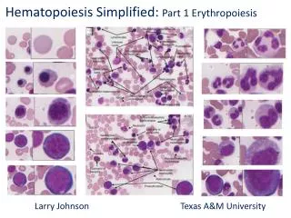
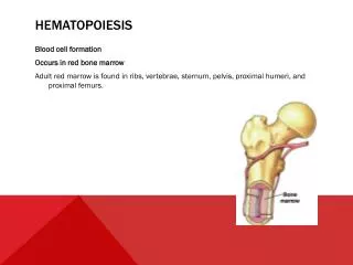
Erythropoiesis Myeloid (blood producing) tissue is found in the red bone marrow located in the spongy bone. As a person ages much of this marrow becomes fatty and ceases production. But it retains stem cells and can be called on to regenerate and produce blood cells later in an emergency. RBCs enter the blood at a rate of about 2 million cells per second.
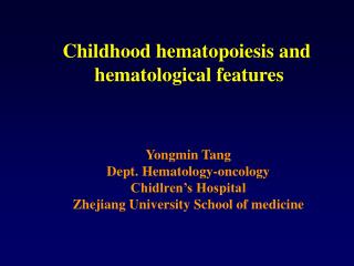
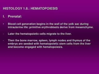
Production of Red Blood Cells Body Areas of the Body That Produce Red Blood Cells. In the early weeks of embryonic life- yolk sac. During the middle trimester of gestation: liver (main), spleen and lymph nodes (additional sites). Then, during the last month of gestation and after birth: exclusively in bone marrow.- Till 5 yrs age: The bone marrow of essentially all bones 5 – 20 years age- The marrow of the long bones, becomes fatty and produces no more red blood cells after about age 20 years. Beyond 20 years, most red cells are produced in the marrow of the membranous bones, such as the vertebrae, sternum, ribs, and ilia.
Genesis of Blood Cells Pluripotential Hematopoietic Stem Cells, Growth Inducers, and Differentiation Inducers. The blood cells begin their lives in the bone marrow from pluripotential hematopoietic stem cell (PHSC). There are successive divisions of the pluripotential cells to form the different circulating blood cells. The intermediate-stage cells are very much like the pluripotential stem cells, but are committed to a particular line of cells and are called committed stem cells.
Anemias Anemia means deficiency of hemoglobin in the blood, which can be caused by: Blood Loss Anemia. After rapid hemorrhage, the body replaces the fluid portion of the plasma in 1 to 3 days, but this leaves a low concentration of red blood cells. The Hb returns to normal by 3 to 6 weeks. In chronic blood loss, a person frequently cannot absorb enough iron from the intestines to form hemoglobin as rapidly as it is lost. Red cells are then produced that are much smaller than normal and have too little hemoglobin inside them, giving rise to microcytic, hypochromic anemia, which is shown in Figure in next slide. 2. Aplastic Anemia. Bone marrow aplasia means lack of functioning bone marrow. For instance, a person exposed to gamma ray radiation from a nuclear bomb blast can sustain complete destruction of bone marrow
3. Megaloblastic Anemia. Deficiency of Vitamin B12, folic acid, and absence of intrinsic factor from the stomach mucosa lead to slow reproduction of erythroblasts in the bone marrow. As a result, the red cells grow too large, with odd shapes, and are called megaloblasts. Thus, atrophy of the stomach mucosa, as occurs in pernicious anemia, or loss of the entire stomach after surgical total gastrectomy can lead to megaloblastic anemia. Also, patients who have intestinal sprue, in which folic acid, vitamin B12, and other vitamin B compounds are poorly absorbed, develop megaloblastic anemia.
4. Hemolytic Anemia. Due to Breakdown of RBCs. Inherited defects of Red cell membrane make the cells fragile, so that they rupture easily as they go through the capillaries, especially through the spleen. The number of red blood cells formed may be normal in hemolytic diseases but the life span of the fragile red cell is so short that the cells are destroyed faster than they can be formed. Eg a. hereditary spherocytosis- the red cells are very small and spherical rather than being biconcave discs. These cells cannot withstand compression forces because they do not have the normal loose, baglike cell membrane structure of the biconcave discs. On passing through the splenic pulp and some other tight vascular beds, they are easily ruptured by even slight compression.
b. Sickle cell anemia- The RBCs have an abnormal type of hemoglobin called hemoglobin S, containing faulty beta chains in the hemoglobin molecule, When this hemoglobin is exposed to low concentrations of oxygen, it precipitates into long crystals inside the red blood cell. These crystals elongate the cell and give it the appearance of a sickle rather than a biconcave disc. The precipitated hemoglobin also damages the cell membrane, so that the cells become highly fragile, leading to serious anemia. Such patients frequently experience a circle of events called a sickle cell disease “sicke cell crisis," in which low oxygen tension in the tissues causes sickling, which leads to ruptured red cells, which causes a further decrease in oxygen tension and still more sickling and red cell destruction. c. Erythroblastosis fetalis, Rh-positive red blood cells in the fetus are attacked by antibodies from an Rh-negative mother. These antibodies make the Rh-positive cells fragile, leading to rapid rupture and causing the child to be born with serious anemia.
Effects of Anemia on Function of the Circulatory System. The blood viscosity falls. This decreases the resistance to blood flow in the peripheral blood vessels, so that far greater than normal quantities of blood flow through the tissues and return to the heart, thereby greatly increasing cardiac output. Moreover, hypoxia resulting from diminished transport of oxygen by the blood causes the peripheral tissue blood vessels to dilate, allowing a further increase in the return of blood to the heart and increasing the cardiac output to a still higher level-sometimes three to four times normal. Thus, one of the major effects of anemia is greatly increased cardiac output, as well as increased pumping workload on the heart.
5. When a person with anemia begins to exercise, the heart is not capable of pumping much greater quantities of blood than it is already pumping. Consequently, during exercise, which greatly increases tissue demand for oxygen, extreme tissue hypoxia results, and acute cardiac failure ensues
EOSINOPHILS 3 – 8% of the Leucocytes. Have a typical ‘Spectacle shaped’, bilobed nucleus. Have coarse bright pink staining granules in the cytoplasm.
LYMPHOCYTES: IMMUNOCYTES Morphologically, LARGE Lymphocytes: Sized about 12 – 15 µ Thin cytoplasmic rim Large spherical nucleus No cytoplasmic granules.
LYMPHOCYTES: IMMUNOCYTES SmallLymphocytes: Sized about 8 µ. ( Smallest Leucocytes) Thin cytoplasmic rim & Large spherical nucleus. No granules visible.
Aplastic Anemia Bone Marrow Failure & ↓ cells production Better called Hypoplastic Anemia ( rarity of total bone marrow aplasia) This leads to failure of production of various cell lineages (Anemia, Leukopenia & Thrombocytopenia)
Etiology I. Hereditary II. Acquired Hereditary – e.g. Fanconi Aplastic Anemia Acquired – Idiopathic -- Radiation Exposure -- Chemicals e.g. Benzene -- Drugs e.g. Chloramphenicol, Cytotoxic drugs, gold salts -- Viruses e.g. Parvovirus B19, EBV, HBV, HIV -- Bone marrow destruction & infiltration e.g. Osteopetrosis -- Paroxysmal Nocturnal Hburia (PNH) -- Conn ts dis e.g. SLE -- Pregnancy
Fanconi Anemia * AR inheritance * 1st described by Fanconi 1927 (3 brothers) * Defect in genes modulating DNA stability * After 5 y age – anemia, bleeding, café au lait skin pigmentation, short stature, microcephaly, hypoplastic thumb, dysplastic radii, mental retardation, hypogonadism
Clinical Features Aplastic An * Gradual onset * Pallor, easy fatigue, SOB, Skin bruises, epistaxis, vaginal bleeding, CNS hg * Fever, chills, pharyngitis * No splenomegaly or LN
DX CBC – Pancytopenia (Anemia, Leukopenia, Thrombocytopenia) -- Low reticulocyte count -- relative lymphocytosis BMA&Biopsy– devoid of active hematopoietic tissue -- replaced by fatty tissue -- lymphocytes seen
DD 1- MDS –hyperplastic BM 2- Acute Leukemia- hyperplastic BM, Blasts 3- PNH – CD59 –ve 4- Hairy cell Leukemia – splenomegaly 5- SLE
Treatment 1- BMT & stem cell T – best ttt child, healthy adults & for severe cases & refractory pt * Matched healthy relative donor 2- Conservative therapy: a) Anti-thymocyte globulin (ATG) 15-40 mg/kg/d for 4 days + Methyl PDN pulse S/E serum sickness, allergy 35 % relapse within 5 years
b) Cyclosporine – cyclic polypeptide inhibiting IL-2 production by T cells - orally 10-12 mg/kg/d for 4-6 months. - S/E hypertension, hyperglycemia, hypertelorism - equally effective as ATG c) Cyclophosphamide –alkylating agent, immunosuppressant - high dose needed - 65% CR
d) Andrgens – not effective in trials e) Cytokines – G-CSF, GM-CSF - not much beneficial 3- Others - IVIG
Prognosis *Before BMT & Immunoth – 25% die in 4 months & 50% in 1 y * BMT cure 80% < 20 y age 70% 20-40y 50% > 40 y