Hematopoiesis
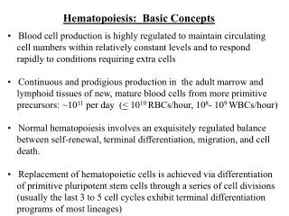
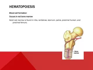
Hematopoiesis. Blood cell formation Occurs in red bone marrow Adult red marrow is found in ribs, vertebrae, sternum, pelvis, proximal humeri, and proximal femurs. Hematopoiesis. All blood cells are derived from a common stem cell ( hemocytoblast ) Hemocytoblast differentiation

Hematopoiesis
E N D
Presentation Transcript
Hematopoiesis • Blood cell formation • Occurs in red bone marrow • Adult red marrow is found in ribs, vertebrae, sternum, pelvis, proximal humeri, and proximal femurs.
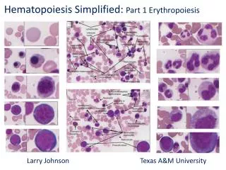
Hematopoiesis • All blood cells are derived from a common stem cell (hemocytoblast) • Hemocytoblast differentiation • Lymphoid stem lymphocytes • Myeloid stem cell all other formed elements Figure 10.4
Formation of Erythrocytes • Mature RBC are anucleate= NO NUCLEUS. Therefore, Unable to divide, grow, or synthesize proteinsWear out in 100 to 120 days • When worn out, RBCs are eliminated by phagocytes in the spleen or liver • Lost cells are replaced by division of hemocytoblasts in the red bone marrow
Stimulus: DecreasedRBC count, decreasedavailability of O2 toblood, or increasedtissue demands for O2 Imbalance Normal blood oxygen levels Imbalance IncreasedO2- carryingability of blood Reduced O2levels in blood MoreRBCs Kidney releaseserythropoietin Enhancederythropoiesis Erythropoietinstimulates Red bonemarrow Control of Erythrocyte Production Rate is controlled by a hormone (erythropoietin) Kidneys produce most erythropoietin as a response to reduced oxygen levels in the blood Homeostasis is maintained by negative feedback from blood oxygen levels Figure 10.5
Normal blood oxygen levels Control of Erythrocyte Production Figure 10.5, step 1
Imbalance Stimulus: DecreasedRBC count, decreasedavailability of O2 toblood, or increasedtissue demands for O2 Normal blood oxygen levels Imbalance Control of Erythrocyte Production Figure 10.5, step 2
Imbalance Stimulus: DecreasedRBC count, decreasedavailability of O2 toblood, or increasedtissue demands for O2 Normal blood oxygen levels Imbalance Reduced O2levels in blood Control of Erythrocyte Production Figure 10.5, step 3
Imbalance Stimulus: DecreasedRBC count, decreasedavailability of O2 toblood, or increasedtissue demands for O2 Normal blood oxygen levels Imbalance Reduced O2levels in blood Kidney releaseserythropoietin Control of Erythrocyte Production Figure 10.5, step 4
Imbalance Stimulus: DecreasedRBC count, decreasedavailability of O2 toblood, or increasedtissue demands for O2 Normal blood oxygen levels Imbalance Reduced O2levels in blood Kidney releaseserythropoietin Erythropoietinstimulates Red bonemarrow Control of Erythrocyte Production Figure 10.5, step 5
Imbalance Stimulus: DecreasedRBC count, decreasedavailability of O2 toblood, or increasedtissue demands for O2 Normal blood oxygen levels Imbalance Reduced O2levels in blood MoreRBCs Kidney releaseserythropoietin Enhancederythropoiesis Erythropoietinstimulates Red bonemarrow Control of Erythrocyte Production Figure 10.5, step 6
Stimulus: DecreasedRBC count, decreasedavailability of O2 toblood, or increasedtissue demands for O2 Normal blood oxygen levels IncreasedO2- carryingability of blood Reduced O2levels in blood MoreRBCs Kidney releaseserythropoietin Enhancederythropoiesis Erythropoietinstimulates Red bonemarrow Control of Erythrocyte Production Figure 10.5, step 7
Imbalance Stimulus: DecreasedRBC count, decreasedavailability of O2 toblood, or increasedtissue demands for O2 Normal blood oxygen levels Imbalance IncreasedO2- carryingability of blood Reduced O2levels in blood MoreRBCs Kidney releaseserythropoietin Enhancederythropoiesis Erythropoietinstimulates Red bonemarrow Control of Erythrocyte Production Figure 10.5, step 8
Formation of White Blood Cells Colony stimulating factors (CSFs) and interleukins prompt bone marrow to generate leukocytes
Formation of Platelets The stem cell (megakaryocyte) undergoes mitosis without cytokinesis many times, forming a large, multinuclear cell, which then fragments into platelets.
Hemostasis • Stoppage of bleeding resulting from a break in a blood vessel • Hemostasis involves three phases • Vascular spasms • Platelet plug formation • Coagulation (blood clotting)
Hemostasis Figure 10.6
Step 1: Vascular Spasms Hemostasis- STEP 1 • Vascular spasms • Vasoconstriction causes blood vessel to spasm • Spasms narrow the blood vessel, decreasing blood loss Figure 10.6, step 1
Step 1: Vascular Spasms Step 2:Platelet Plug Formation Injury to liningof vessel exposescollagen fibers;platelets adhere Collagenfibers Hemostasis- Step 2 • Platelet plug formation • Collagen fibers are exposed by a break in a blood vessel • Platelets become “sticky” and cling to fibers • Anchored platelets release chemicals to attract more platelets • Platelets pile up to form a platelet plug Figure 10.6, step 2
Step 1: Vascular Spasms Step 2:Platelet Plug Formation Injury to liningof vessel exposescollagen fibers;platelets adhere Plateletplugforms Collagenfibers Platelets Platelets release chemicalsthat attract more platelets tothe site and make nearbyplatelets sticky PF3 fromplatelets Calciumand otherclottingfactorsin bloodplasma + Tissue factorin damagedtissue Hemostasis – step 3 • Coagulation • Injured tissues release tissue factor (TF) • PF3 (a phospholipid) interacts with TF, blood protein clotting factors, and calcium ions to trigger a clotting cascade • Prothrombin activator converts prothrombin to thrombin (an enzyme) Figure 10.6, step 4
Platelets release chemicalsthat attract more platelets tothe site and make nearbyplatelets sticky PF3 fromplatelets Calciumand otherclottingfactorsin bloodplasma + Tissue factorin damagedtissue Phases ofcoagulation(clottingcascade) Formation ofprothrombinactivator Prothrombin Thrombin Fibrinogen(soluble) Fibrin(insoluble) Hemostasis – coagulation continued • Thrombin joins fibrinogen proteins into hair-like molecules of insoluble fibrin • Fibrin forms a meshwork (the basis for a clot) Figure 10.6, step 7
Hemostasis • Blood usually clots within 3 to 6 minutes • The clot remains as endothelium regenerates • The clot is broken down after tissue repair Figure 10.7
Summary of hemostasis • Hemostasis is initiated by a break in the blood vessel wall (or lining), initiating vascular spasms and causing platelets to cling to the damaged site. Once attached, the platelets release serotonin, which enhances vasoconstriction. • Injured tissue cells release thromboplastin, which interacts with platelet phospholipids (PF3), Ca2+ and plasma clotting factors to form prothrombin activator. • Prothrombin activator converts prothrombin to thrombin. Thrombin, an enzyme, then converts soluble fibrinogen molecules into long fibrin threads, which form the basis of the clot.
Undesirable Clotting • Thrombus • A clot in an unbroken blood vessel • Can be deadly in areas like the heart • Embolus • A thrombus that breaks away and floats freely in the bloodstream • Can later clog vessels in critical areas such as the brain Coagulation can be promoted by: i. Roughened vessel lining, which attracts/activates platelets. ii. Pooling of blood within vessels can result in the activation of clotting factors and the initiation of the coagulation process.
Bleeding Disorders • Thrombocytopenia • Platelet deficiency • Even normal movements can cause bleeding from small blood vessels that require platelets for clotting • The liver is the source of fibrinogen and several other factors that are necessary for clotting. When the liver is damaged and dysfunctional, it becomes unable to synthesize the usual amounts of clotting factors. When this situation happens, abnormal and often severe bleeding episodes can occur • Hemophilia • Hereditary bleeding disorder • Normal clotting factors are missing • http://www.sciencecases.org/hemo/hemo.asp
Review Questions #1 • What is the name of the stem cell that gives rise to all other formed elements? • Name the formed elements that arise from myeloid stem cells. Name those arising from lymphoid stem cells. • What property of RBCs dooms them to a limited life span of only 120 days? • What WBC type resides primarily in the tissues of the body? • How is the production of platelets different from that of all other formed elements? • Describe the process of hemostasis. Indicate what starts the process. • What factors enhance the risk of thrombus formation in intact blood vessels? • How can liver dysfunction cause bleeding disorders?
Blood Groups and Transfusions • Large losses of blood have serious consequences • Loss of 15–30% causes weakness • Loss of over 30% causes shock, which can be fatal • Transfusions are the only way to replace blood quickly • Transfused blood must be of the same blood group
Human Blood Groups • Blood contains genetically determined proteins • Antigens (a substance the body recognizes as foreign) may be attacked by the immune system • Antibodies are the “recognizers” • Blood is “typed” by using antibodies that will cause blood with certain proteins to clump (agglutination)
Human Blood Groups • There are over 30 common red blood cell antigens • The most vigorous transfusion reactions are caused by ABO and Rh blood group antigens
ABO Blood Groups • Based on the presence or absence of two antigens • Type A • Type B • The lack of these antigens is called type O
ABO Blood Groups • The presence of both antigens A and B is called type AB • The presence of antigen A is called type A • The presence of antigen B is called type B • The lack of both antigens A and B is called type O
ABO Blood Groups • Blood type AB can receive A, B, AB, and O blood • Universal recipient • Blood type B can receive B and O blood • Blood type A can receive A and O blood • Blood type O can receive O blood • Universal donor
ABO Blood Groups Table 10.3
Rh Blood Groups • Named because of the presence or absence of one of eight Rh antigens (agglutinogen D) that was originally defined in Rhesus monkeys • Most Americans are Rh+ (Rh positive) • Problems can occur in mixing Rh+ blood into a body with Rh– (Rh negative) blood • Percentage of Population with Each Blood Type
Rh Dangers During Pregnancy • Danger occurs only when the mother is Rh– and the father is Rh+, and the child inherits the Rh+ factor • RhoGAM shot can prevent buildup of anti-Rh+ antibodies in mother’s blood
Rh Dangers During Pregnancy • The mismatch of an Rh– mother carrying an Rh+ baby can cause problems for the unborn child • The first pregnancy usually proceeds without problems • The immune system is sensitized after the first pregnancy • In a second pregnancy, the mother’s immune system produces antibodies to attack the Rh+ blood (hemolytic disease of the newborn)
Blood Typing • Blood samples are mixed with anti-A and anti-B serum • Coagulation or no coagulation leads to determining blood type • Typing for ABO and Rh factors is done in the same manner • Cross matching—testing for agglutination of donor RBCs by the recipient’s serum, and vice versa Figure 10.8
Review Questions #2 • What are the classes of human blood groups based on? • What are agglutinins? • Name the four ABO blood groups. • What is a transfusion reaction? Why does it happen? • 10. What is the probable result from infusion of mismatched blood? • 11. Cary is bleeding profusely after being hit by a truck as he was pedaling his bike home. At the hospital, the nurse asked him whether he knew his blood type. He told her he “had the same blood as most other people.” What is his ABO blood type? • What is the difference between an antigen and an antibody? • Explain why a Rh- person does not have a transfusion reaction on the first exposure to Rh+ blood? Why is there a transfusion reaction the second time he or she receives the Rh+ blood?