17
17. Blood. Blood Composition. Blood: a fluid connective tissue composed of Plasma Formed elements Erythrocytes (red blood cells, or RBCs) Leukocytes (white blood cells, or WBCs) Platelets. Blood Composition. Hematocrit Percent of blood volume that is RBCs 47% ± 5% for males

17
E N D
Presentation Transcript
17 Blood
Blood Composition • Blood: a fluid connective tissue composed of • Plasma • Formed elements • Erythrocytes (red blood cells, or RBCs) • Leukocytes (white blood cells, or WBCs) • Platelets
Blood Composition • Hematocrit • Percent of blood volume that is RBCs • 47% ± 5% for males • 42% ± 5% for females
Formed elements Plasma • 55% of whole blood • Least dense component Buffy coat • Leukocytes and platelets • <1% of whole blood Erythrocytes 2 • 45% of whole blood • Most dense component 1 Centrifuge the blood sample. Withdraw blood and place in tube. Figure 17.1
Physical Characteristics and Volume • Sticky, opaque fluid • Color scarlet to dark red • pH 7.35–7.45 • 38C • ~8% of body weight • Average volume: 5–6 L for males, and 4–5 L for females
Functions of Blood • Distribution of • O2 and nutrients to body cells • Metabolic wastes to the lungs and kidneys for elimination • Hormones from endocrine organs to target organs
Functions of Blood • Regulation of • Body temperature by absorbing and distributing heat • Normal pH using buffers • Adequate fluid volume in the circulatory system
Functions of Blood • Protection against • Blood loss • Plasma proteins and platelets initiate clot formation • Infection • Antibodies • Complement proteins • WBCs defend against foreign invaders
Blood Plasma • 90% water • Proteins are mostly produced by the liver • 60% albumin • 36% globulins • 4% fibrinogen
Blood Plasma • Nitrogenous by-products of metabolism—lactic acid, urea, creatinine • Nutrients—glucose, carbohydrates, amino acids • Electrolytes—Na+, K+, Ca2+, Cl–, HCO3– • Respiratory gases—O2 and CO2 • Hormones
Formed Elements • Only WBCs are complete cells • RBCs have no nuclei or organelles • Platelets are cell fragments • Most formed elements survive in the bloodstream for only a few days • Most blood cells originate in bone marrow and do not divide
Platelets Erythrocytes Monocyte Neutrophils Lymphocyte Figure 17.2
Erythrocytes • Biconcave discs, anucleate, essentially no organelles • Filled with hemoglobin (Hb) for gas transport • Contain the plasma membrane protein spectrin and other proteins • Provide flexibility to change shape as necessary • Are the major factor contributing to blood viscosity
2.5 µm Side view (cut) 7.5 µm Top view Figure 17.3
Erythrocytes • Structural characteristics contribute to gas transport • Biconcave shape—huge surface area relative to volume • >97% hemoglobin (not counting water) • No mitochondria; ATP production is anaerobic; no O2 is used in generation of ATP • A superb example of complementarity of structure and function!
Erythrocyte Function • RBCs are dedicated to respiratory gas transport • Hemoglobin binds reversibly with oxygen
Erythrocyte Function • Hemoglobin structure • Protein globin: two alpha and two beta chains • Heme pigment bonded to each globin chain • Iron atom in each heme can bind to one O2 molecule • Each Hb molecule can transport four O2
bGlobin chains Heme group a Globin chains (a) Hemoglobin consists of globin (two alpha and two beta polypeptide chains) and four heme groups. (b) Iron-containing heme pigment. Figure 17.4
Hemoglobin (Hb) • O2 loading in the lungs • Produces oxyhemoglobin (ruby red) • O2 unloading in the tissues • Produces deoxyhemoglobin or reduced hemoglobin (dark red) • CO2 loading in the tissues • Produces carbaminohemoglobin (carries 20% of CO2 in the blood)
Hematopoiesis • Hematopoiesis (hemopoiesis): blood cell formation • Occurs in red bone marrow of axial skeleton, girdles and proximal epiphyses of humerus and femur
Hematopoiesis • Hemocytoblasts (hematopoietic stem cells) • Give rise to all formed elements • Hormones and growth factors push the cell toward a specific pathway of blood cell development • New blood cells enter blood sinusoids
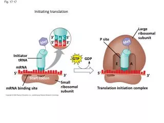
Erythropoiesis • Erythropoiesis: red blood cell production • A hemocytoblast is transformed into a proerythroblast • Proerythroblasts develop into early erythroblasts
Erythropoiesis • Phases in development • Ribosome synthesis • Hemoglobin accumulation • Ejection of the nucleus and formation of reticulocytes • Reticulocytes then become mature erythrocytes
Stem cell Committed cell Developmental pathway Phase 1 Ribosome synthesis Phase 2 Hemoglobin accumulation Phase 3 Ejection of nucleus Reticulo- cyte Erythro- cyte Proerythro- blast Early erythroblast Late erythroblast Normoblast Hemocytoblast Figure 17.5
Regulation of Erythropoiesis • Too few RBCs leads to tissue hypoxia • Too many RBCs increases blood viscosity • Balance between RBC production and destruction depends on • Hormonal controls • Adequate supplies of iron, amino acids, and B vitamins
Hormonal Control of Erythropoiesis • Erythropoietin (EPO) • Direct stimulus for erythropoiesis • Released by the kidneys in response to hypoxia
Hormonal Control of Erythropoiesis • Causes of hypoxia • Hemorrhage or increased RBC destruction reduces RBC numbers • Insufficient hemoglobin (e.g., iron deficiency) • Reduced availability of O2 (e.g., high altitudes)
Hormonal Control of Erythropoiesis • Effects of EPO • More rapid maturation of committed bone marrow cells • Increased circulating reticulocyte count in 1–2 days • Testosterone also enhances EPO production, resulting in higher RBC counts in males
IMBALANCE Homeostasis: Normal blood oxygen levels 1 Stimulus: Hypoxia (low blood O2- carrying ability) due to • DecreasedRBC count • Decreased amountof hemoglobin • Decreasedavailability of O2 5 O2- carryingability of bloodincreases. IMBALANCE 4 Enhancederythropoiesisincreases RBCcount. 2 Kidney (and liver toa smaller extent)releaseserythropoietin. 3 Erythropoietinstimulates redbone marrow. Figure 17.6
IMBALANCE Homeostasis: Normal blood oxygen levels 1 Stimulus: Hypoxia (low blood O2- carrying ability) due to • DecreasedRBC count • Decreased amountof hemoglobin • Decreasedavailability of O2 IMBALANCE Figure 17.6, step 1
IMBALANCE Homeostasis: Normal blood oxygen levels 1 Stimulus: Hypoxia (low blood O2- carrying ability) due to • DecreasedRBC count • Decreased amountof hemoglobin • Decreasedavailability of O2 IMBALANCE 2 Kidney (and liver toa smaller extent)releaseserythropoietin. Figure 17.6, step 2
IMBALANCE Homeostasis: Normal blood oxygen levels 1 Stimulus: Hypoxia (low blood O2- carrying ability) due to • DecreasedRBC count • Decreased amountof hemoglobin • Decreasedavailability of O2 IMBALANCE 2 Kidney (and liver toa smaller extent)releaseserythropoietin. 3 Erythropoietinstimulates redbone marrow. Figure 17.6, step 3
IMBALANCE Homeostasis: Normal blood oxygen levels 1 Stimulus: Hypoxia (low blood O2- carrying ability) due to • DecreasedRBC count • Decreased amountof hemoglobin • Decreasedavailability of O2 IMBALANCE 4 Enhancederythropoiesisincreases RBCcount. 2 Kidney (and liver toa smaller extent)releaseserythropoietin. 3 Erythropoietinstimulates redbone marrow. Figure 17.6, step 4
IMBALANCE Homeostasis: Normal blood oxygen levels 1 Stimulus: Hypoxia (low blood O2- carrying ability) due to • DecreasedRBC count • Decreased amountof hemoglobin • Decreasedavailability of O2 5 O2- carryingability of bloodincreases. IMBALANCE 4 Enhancederythropoiesisincreases RBCcount. 2 Kidney (and liver toa smaller extent)releaseserythropoietin. 3 Erythropoietinstimulates redbone marrow. Figure 17.6, step 5
Dietary Requirements for Erythropoiesis • Nutrients—amino acids, lipids, and carbohydrates • Iron • Stored in Hb (65%), the liver, spleen, and bone marrow • Stored in cells as ferritin and hemosiderin • Transported loosely bound to the protein transferrin • Vitamin B12 and folic acid—necessary for DNA synthesis for cell division
Fate and Destruction of Erythrocytes • Life span: 100–120 days • Old RBCs become fragile, and Hb begins to degenerate • Macrophages engulf dying RBCs in the spleen
Fate and Destruction of Erythrocytes • Heme and globin are separated • Iron is salvaged for reuse • Heme is degraded to yellow the pigment bilirubin • Liver secretes bilirubin (in bile)) into the intestines • Degraded pigment leaves the body in feces as stercobilin • Globin is metabolized into amino acids
1 Low O2levels in blood stimulate kidneys to produce erythropoietin. 2 Erythropoietin levels rise in blood. 3 Erythropoietin and necessary raw materials in blood promote erythropoiesis in red bone marrow. 4 New erythrocytes enter bloodstream; function about 120 days. 5 Aged and damaged red blood cells are engulfed by macrophages of liver, spleen, and bone marrow; the hemoglobin is broken down. Hemoglobin Heme Globin Bilirubin Amino acids Iron stored as ferritin, hemosiderin Iron is bound to transferrin and released to blood from liver as needed for erythropoiesis. Bilirubin is picked up from blood by liver, secreted into intestine in bile, metabolized to stercobilin by bacteria, and excreted in feces. Circulation Food nutrients, including amino acids, Fe, B12, and folic acid, are absorbed from intestine and enter blood. 6 Raw materials are made available in blood for erythrocyte synthesis. Figure 17.7
1 Low O2levels in blood stimulate kidneys to produce erythropoietin. Figure 17.7, step 1
1 Low O2levels in blood stimulate kidneys to produce erythropoietin. 2 Erythropoietin levels rise in blood. Figure 17.7, step 2
1 Low O2levels in blood stimulate kidneys to produce erythropoietin. 2 Erythropoietin levels rise in blood. 3 Erythropoietin and necessary raw materials in blood promote erythropoiesis in red bone marrow. Figure 17.7, step 3
1 Low O2levels in blood stimulate kidneys to produce erythropoietin. 2 Erythropoietin levels rise in blood. 3 Erythropoietin and necessary raw materials in blood promote erythropoiesis in red bone marrow. 4 New erythrocytes enter bloodstream; function about 120 days. Figure 17.7, step 4
Hemoglobin 5 Aged and damaged red blood cells are engulfed by macrophages of liver, spleen, and bone marrow; the hemoglobin is broken down. Heme Globin Bilirubin Amino acids Iron stored as ferritin, hemosiderin Bilirubin is picked up from blood by liver, secreted into intestine in bile, metabolized to stercobilin by bacteria, and excreted in feces. Circulation Figure 17.7, step 5
Hemoglobin 5 Aged and damaged red blood cells are engulfed by macrophages of liver, spleen, and bone marrow; the hemoglobin is broken down. Heme Globin Bilirubin Amino acids Iron stored as ferritin, hemosiderin Iron is bound to transferrin and released to blood from liver as needed for erythropoiesis. Bilirubin is picked up from blood by liver, secreted into intestine in bile, metabolized to stercobilin by bacteria, and excreted in feces. Circulation Food nutrients, including amino acids, Fe, B12, and folic acid, are absorbed from intestine and enter blood. 6 Raw materials are made available in blood for erythrocyte synthesis. Figure 17.7, step 6
1 Low O2levels in blood stimulate kidneys to produce erythropoietin. 2 Erythropoietin levels rise in blood. 3 Erythropoietin and necessary raw materials in blood promote erythropoiesis in red bone marrow. 4 New erythrocytes enter bloodstream; function about 120 days. 5 Aged and damaged red blood cells are engulfed by macrophages of liver, spleen, and bone marrow; the hemoglobin is broken down. Hemoglobin Heme Globin Bilirubin Amino acids Iron stored as ferritin, hemosiderin Iron is bound to transferrin and released to blood from liver as needed for erythropoiesis. Bilirubin is picked up from blood by liver, secreted into intestine in bile, metabolized to stercobilin by bacteria, and excreted in feces. Circulation Food nutrients, including amino acids, Fe, B12, and folic acid, are absorbed from intestine and enter blood. 6 Raw materials are made available in blood for erythrocyte synthesis. Figure 17.7
Erythrocyte Disorders • Anemia: blood has abnormally low O2-carrying capacity • A sign rather than a disease itself • Blood O2 levels cannot support normal metabolism • Accompanied by fatigue, paleness, shortness of breath, and chills
Causes of Anemia • Insufficient erythrocytes • Hemorrhagic anemia: acute or chronic loss of blood • Hemolytic anemia: RBCs rupture prematurely • Aplastic anemia: destruction or inhibition of red bone marrow
Causes of Anemia • Low hemoglobin content • Iron-deficiency anemia • Secondary result of hemorrhagic anemia or • Inadequate intake of iron-containing foods or • Impaired iron absorption
Causes of Anemia • Pernicious anemia • Deficiency of vitamin B12 • Lack of intrinsic factor needed for absorption of B12 • Treated by intramuscular injection of B12 or application of Nascobal
Causes of Anemia • Abnormal hemoglobin • Thalassemias • Absent or faulty globin chain • RBCs are thin, delicate, and deficient in hemoglobin