Unit 7 Rh BLOOD GROUP SYSTEM
Unit 7 Rh BLOOD GROUP SYSTEM. Terry Kotrla, MS, MT(ASCP)BB. Introduction. Rh is the most important blood group system after ABO in transfusion medicine. One of the most complex of all RBC blood group systems with more than 50 different Rh antigens.

Unit 7 Rh BLOOD GROUP SYSTEM
E N D
Presentation Transcript
Unit 7 Rh BLOOD GROUP SYSTEM Terry Kotrla, MS, MT(ASCP)BB
Introduction • Rh is the most important blood group system after ABO in transfusion medicine. • One of the most complex of all RBC blood group systems with more than 50 different Rh antigens. • The genetics, nomenclature and antigenic interactions are unsettled. • This unit will concentrate on the most COMMONLY encountered observations, problems and solutions.
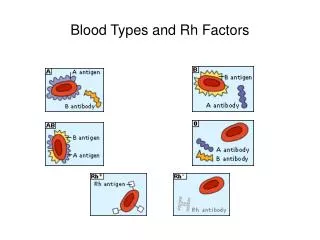
Antigens of Rh System • Terms “D positive” and “D negative” refer only to presence or absence of the Rh antigen D on the red blood cell. • Terms “Rh pos” and “Rhneg” are old terms, although blood products still labeled as such. • Early name “Rho” less frequently used. • Four additional antigens: C, c, E, e. • Named by Fisher for next letters of alphabet according to precedent set by naming A and B blood groups. • Major alleles are C/c and E/e. • MANY variations and combinations of the 5 principle genes and their products, antigens, have been recognized. • The Rh antigens and corresponding antibodies account for majority of unexpected antibodies encountered. • Rh antibodies stimulated as a result of transfusion or pregnancy, they are immune.
HISTORY • Key observation by Levine and Stetson in 1939 that delivery of stillborn fetus and adverse reaction in mom to blood transfusion from father were related. • Syndrome in fetus is now referred to as hemolytic disease of the fetus and newborn (HDFN). • Syndrome had complicated pregnancies for decades causing severe jaundice and fetal death, “erythroblastosisfetalis”. • Erythroblastosisfetalis (HDN) linked with Anti-Rh by Levine in 1941. • Rh system IDENTIFIED by Landsteiner and Wiener in 1940. • Immunized animals to Rhesus macaque monkey RBCs. • Antibody agglutinated 100% of Rhesus and 85% of human RBCs. • Reactivity paralleled reactivity of sera in women who delivered infant suffering from hemolytic disease. • Later antigen detected by rhesus antibody and human antibody established to be dissimilar but system already named.
Clinical Significance • D antigen, after A and B, is the most important RBC antigen in transfusion practice. • Individuals who lack D antigen DO NOT have anti-D. • Antibody produced through exposure to D antigen through transfusion or pregnancy. • Immunogenicity of D greater than that of all other RBC antigens studied. • Has been reported that 80%> of D neg individuals who receive single unit of D pos blood can be expected to develop immune anti-D. • Testing for D is routinely performed so D neg will be transfused with D neg.
Inheritance and Nomenclature • Two systems of nomenclature developed prior to advances in molecular genetics. • Reflect serologic observations and inheritance theories based on family studies. • Because these are used interchangeably it is necessary to understand the theories well enough to translate from one to the other. • Two additional systems developed so universal language available for use with computers.
Fisher-Race: CDE Terminology • Fisher Race • Suggested that antigens are determined by 3 pairs of genes which occupy closely linked loci. • Each gene complex carries D or its absence (d), C or c, E or e. • Each gene (except d, which is an amorph) causes production of an antigen. • The order of loci on the gene appears to be “DCE” but many authors prefer to use “CDE” to follow alphabet. • Inherited from parents in linked fashion as haplotypes • The gene d is assumed to be present when D is absent.
Fisher-Race • Three loci carry the Rh genes are so closely linked that they never separate but are passed from generation to generation as a unit or gene complex.
Fisher-Race • Below an offspring of the Dce/dce individual will inherit EITHER Dce or dce from the parent, never dCe as this would indicate crossing over which does not occur in Rh system in man.
Fisher-Race • With the exception of d each allelic gene controls presence of respective antigen on RBC. • The gene complex DCe would cause production of the D, C and e antigens on the red cells. • If the same gene complex were on both paired chromsomes (DCe/DCe) then only D, C and e would be present on the cells. • If one chromsome carried DCe and the other was DcE this would cause D, C, c, E and e antigens to be present on red blood cells. • Each antigen except d is recognizable by testing red cells with specific antiserum.
Wiener • Postulated that TWO genes, one on each chromosome pair, controls the entire express of Rh system. • Each gene produces a structure on the red cell called an agglutinogen (antigen). • Eight (8) major alleles (agglutinogens): R0, R1, R2, Rz, r, r’, r” and ry. • Each agglutinogen has 3 factors (antigens or epitopes) • The three factors are the antigens expressed on the cell. • For example the agglutinogen R0= Rh0 (D), hr’ (c), hr’ (e) • Each agglutinogen can be identified by its parts or factors that react with specific antibodies (antiserums).
Weiner and Fisher-Race • The two theories are the basis for the two notations currently used for the Rh system. • Immunohematologists use combinations of both systems when recording most probable genotypes. • You MUST be able to convert a Fisher-Race notation into Wiener shorthand, i.e., Dce (Fisher-Race) is written R0. • Given an individual’s phenotype you MUST determine all probable genotypes and write them in both Fisher-Race and Wiener notations. • R1r is the most common D positive genotype. • rr is the most common D negative genotype.
Weiner and Fisher-Race D = R 1 ( C) Z (both C & E ) 2 ( E ) 0 (neither C or E ) D c e D C E D C D c E d = r ‘( C) y (both C & E ) ‘’ ( E ) (neither C or E ) d c e d C E d C e d cE
Differentiating Superscript from Subscript Superscripts (Rh1) refer to genes Subscripts (Rh1) refer to the agglutinogen (complex of antigens) For example, the Rh1 gene codes for the Rh1 agglutinogen made of D, C, e Usually, this can be written in shorthand, leaving out the “h” DCe is written as R1
Converting Wiener into Fisher-Race or Vice Versa R D r no D 1 and ‘ C 2 and “ E Example: DcE R2 r” dcE Written in shorthand
Rosenfield • In 1962 proposed a nomenclature based ONLY on serologic (agglutination) reactions. • Antigens are numbered in the order of their discovery and recognition as belonging to the Rh system. • No genetic assumptions made • The phenotype of a given cell is expressed by the base symbol of “Rh” followed by a colon and a list of the numbers of the specific antisera used. • If listed alone, the Antigen is present (Rh:1 = D Ag) • If listed with a “-”, antigen is not present (Rh:1, -2, 3 = DcE) • If not listed, the antigen status was not determined • Adapts well to computer entry
International Society of Blood Transfusion • Abbreviated ISBT • International organization created to standardize blood group system nomenclature. • Assigned 6 digit number for each antigen. • First 3 numbers indicate the blood group system, eg., 004 = Rh • Last 3 numbers indicates the specific antigen, eg., 004001 = D antigen. • For recording of phenotypes, the system adopts the Rosenfield approach
Phenotype versus Genotype The phenotype is the result of the reaction between the red cells and antisera The genotype is the genetic makeup and can be predicted using the phenotype and by considering the race of an individual Only family studies can determine the true genotype
Five reagent antisera available. Only anti-D required for routine testing. Other typing sera used for typing rbcs to resolve antibody problems or conduct family studies. Agglutination reactions (positive and negative) will represent the phenotype. No anti-d since d is an amorph. Use statistical probability to determine most probable genotype. Phenotyping and Genotyping
Rh Phenotyping • Uses • Parentage testing • Predicting hemolytic disease of the fetus and newborn (HDFN) • Confirmation of Rh antibody specificity • Locating compatible blood for recipients with Rh antibodies. • Protocol • Mix unknown RBCs with Rh antisera • Agglutination indicates presence of antigen on cell and determines phenotype. • Use published frequencies and subject information to determine genotype.
Phenotyping and Genotyping Molecular testing becoming more popular: Cannot use anti-sera on recently transfused individuals, molecular testing can differentiate. Anti-sera not available for some antigens, molecular testing being developed for all blood group genes. D zygosity can be determined. Fetal genotyping for D can be done on fetal DNA present in maternal plasma. Monoclonal reagents from different manufacturers react differently with variant D antigens, molecular test specific. Typing sera continue to be the “gold standard” but this will change in the future.
Genotype Frequencies • Refer to textbook. • Genotypes are listed as “presumptive” or “most probable”. • Genotypes will vary in frequency in different racial groups.
Weak Expression of D Not all D positive cells react equally well with anti-D. RBCs not immediately agglutinated by anti-D must be tested for weak D. Incubate cells with anti-D at 37C, coating of D antigens will occur if present. Wash X3 add AHG AHG will bind to anti-D coating cells if present. If negative, individual is D negative If positive, individual is D positive w
Three Mechanisms for Weak D Genetic Position effect Mosaic Results in differences from normal D expression Quantitative (inherited weak D or position effects) Qualitative (mosaic D; could produce Anti-D)
Weak D - Genetic Inheritance of D genes which result in lowered densities of D Antigens on RBC membranes, gene codes for less D.
Weak D - Genetic RBC with normal amounts of D antigen Weak D (Du)
Position Effect C trans - position effect; The D gene is in trans to the C gene, eg., C and D are on OPPOSITE sides: Dce/dCe C and D antigen arrangement causes steric hindrance which results in weakening or suppression of D expression.
Position Effect C in trans position to D: D c e / d C e Weak D C in cis position to D: D C e / d c e NOweak D
Partial D Absence of a portion or portions of the total material that comprises the D antigen. Known as “partial D” (old term “D mosaic”).
D Mosaic/Partial D If the patient is transfused with D positive red cells, they may develop an anti-Dalloantibody* to the part of the antigen (epitope) that is missing Missing portion RBC RBC *alloantibody- antibody produced with specificity other than self
Significance of Weak D Donors Labeled as D positive Weak D substantially less immunogenic than normal D Weak D has caused severe HTR in patient with anti-D Patients If weak D due to partial D can make antibody to portion they lack. If weak D due to suppression or genetic expression theoretically could give D positive Standard practice to transfuse with D negative Weak D testing on donors by transfusion service not required. Weak D testing on patients not required except in certain situations.
Compound Antigens Compound antigens are epitopes which occur due to presence of two Rh genes on the same chromosome, cis position. Gene products include not only products of single gene but also a combined gene that is also antigenic. (f, rh1, etc) f antigens occur when c and e are found in cis (Example: dce/dce) r(cde) gene makes c and e but also makes f (ce). ONLY OCCURS when c and e are in the CIS position. f antigen will NOT be present in trans position. rh1 or Ce antigens occur when C and e are in cis (example: dCe/dce) Antibodies rarely encountered but if individual had anti-f would only react with f positive cells, not cells positive for c or e in trans only. f cells clearly marked on antigram of screen and panel cells.
G Antigen Genes that code for C or D also code for G G almost invariably present on RBCs possessing C or D Anti-G mimics anti-C and anti-D. Anti-G activity cannot be separated into anti-C and anti-D.
D Deletion Very rare Individuals inherit Rh gene complex lacking alleles. May be at Ee or Cc Must be homozygous for rare deletion to be detected. No reaction when RBCs are tested with anti-E, anti-e, anti-C or anti-c Requires transfusion of other D-deletion red cells, because these individuals may produce antibodies with single or separate specificities. Written as D- - or -D-
Rh Null Red cells have no Rh antigen sites Genotype written ---/--- The lack of antigens causes the red cell membrane to appear abnormal leading to: Stomatocytosis Hemolytic anemia 2 Rh null phenotypes: Regulator type – gene inherited, but not expressed Amorph type – RHD gene is absent, no expression of RHCE gene Complex antibodies may be produced requiring use of rare, autologous or compatible blood from siblings.
LW Discovered at same time as Rh antigen. LW detected on cells of Rhesus monkeys and human rbcs in same proportion as D antigen. Thought was the same antigen but discovered differences. Named LW in honor of Landsteiner and Wiener. Rare individuals lack LW yet have normal Rh antigens. Can form allo anti-LW. Reacts more strongly with D pos than D neg cells. Keep in mind when D pos individual appears to have anti-D
Cw Variant Rh antigen Low frequency antigen found in only 1-2% of Whites and rare in Blacks Most individuals who are C+ are Cw+ Antibodies to these antigens can be naturally occuring and may play a role in HFDN and HTR
Rh Antibodies Except for rare examples of anti-E and anti-Cw which may be naturally occurring, most occur from immunization due to transfusion or pregnancy. Associated with HTR and HDFN. Characteristics IgG but may have MINOR IgM component so will NOT react in saline suspended cells (IS). May be detected at 37C but most frequently detected by IAT. Enhanced by testing with enzyme treated cells. Order of immunogenicity: D > c > E > C > e Do not bind complement, extravascular destruction.
Rh Antibodies Anti-E most frequently encountered antibody followed by anti-c. Anti-C rare as single antibody. Anti-e rarely encountered as only 2% of the population is antigen negative. Detectable antibody persists for many years and sometimes for life. Anti-D may react more strongly with R2R2 cells than R1R1 due to higher density of D antigen on cells.
Concomitant Rh Antibodies Antibodies which often occur TOGETHER. Sera containing anti-D may contain anti-G (anti-C + -D) Anti-C rarely occurs only, most often with anti-D. Anti-ce (-f) often seen in combinatiion with anti-c. MOST IMPORTANT is R1R1 who make anti-E frequently make anti-c. Patients with anti-E should be phenotyped for c antigen. If patient appears to be R1R1 should be transfused with R1R1 blood. Anti-c frequently falls below detectable levels.
Detection of D Antigens • Four types of anti-D reagents • High Protein - Faster, increased frequency of false positives; requires use of Rh control tube, converts to weak D testing • IgM (Low protein/Saline reacting) - Low protein (fewer false positives); long incubation times; cannot convert to weak D testing • Chemically modified - “Relaxed” form of IgG Anti-D in low protein medium; few false positives; saline control performed; converts to weak D testing • Monoclonal source, low protein, blends of mAbs • Must know the preparation, use, advantages and limitations of each.
High Protein Anti-D IgG anti-D potentiated with high protein and other macromolecules to ensure agglutination at IS. May cause false positives with rbcs coated with antibody. Diluent control REQUIRED. False positives due to autoagglutinins, abnormal serum proteins, antibodies to additives and using unwashed rbcs. Can be used for weak D test.
IgM Anti-D (low protein/saline) Prepared from predominantly IgM antibodies, scarce due to difficulty obtaining raw material. Reserved for individuals giving false positive with high protein anti-seras. Newer saline anti-sera require incubation at 37. No negative control required unless AB positive. CANNOT be used by slide test OR weak D test.
Chemically Modified IgG converted to saline agglutinin by weakining disulfide bonds at hinge region, greater flexibility, increases span distance. Stronger reactivity than IgM antibodies. Can be used for slide, tube and weak D test. Negative control unnecessary unless AB positive.
Monoclonal Anti-D Prepared from blend of moncolonal IgM and polyclonal IgG. IgM reacts at IS IgG reacts at AHG (weak D test) Most frequently utilized reagent. Used for tube, slide and weak D test. Negative control unnecessary unless AB positive.
Control for Low Protein Reagents Diluent used has protein concentration equaling human serum. False positives due to immunoglobulin coating of test rbcs occurs no more frequently than with other saline reactive anti-sera. False positives do occur, patient will appear to be AB positive on forward type. Must run saline or manufacturer’s control to verify.
Precautions for Rh Typing MUST follow manufacturer’s instructions as testing protocols vary. Cannot use IAT unless explicitly instructed by manufacturer. Positive and negative controls must be tested in parallel with test rbcs. QC performed daily for anti-D QC for other anti-seras performed in parallel with test since these are usually not tested each day, only when necessary.