cerebral circulation & CSF
cerebral circulation & CSF. Sundaresh T. V. Department of Neurosciences. Windsor University. St Kitts. Blood Supply of Brain & Applied aspects. internal carotid circulation V ertebrobasilar circulation Circle of Willis ‘Stroke’ Venous drainage CSF. Internal carotid artery.

cerebral circulation & CSF
E N D
Presentation Transcript
cerebral circulation & CSF Sundaresh T. V. Department of Neurosciences. Windsor University. St Kitts.
Blood Supply of Brain & Applied aspects • internal carotid circulation • Vertebrobasilar circulation • Circle of Willis • ‘Stroke’ • Venous drainage • CSF
Internal carotid artery • Arises from common carotid artery • Enters skull through carotid canal of petrous bone & proceeds through cavernous sinus curving in an ‘S’ shape (carotid siphon) • Supplies rostral 2/3rds of brain including main parts of basal nuclei & internal capsule
Internal carotid artery • gives off several collateral branches before it divides into its two terminal branches: • middle cerebral artery & • anterior cerebral artery
ICA - collateral branches • inferior & superior hypophyseal arteries & the ophthalmic artery come off the ica as soon as the artery has entered the skull • posterior communicating & anterior choroidal arteries are the other named branches
Ophthalmic artery 1. ethmoidal arteries 2. ciliary arteries 3. optic nerve & ophthalmic artery 4. central retinal artery 5. retinal arteries 6. supratrochlear artery 7. supraorbital artery 8. dorsal nasal artery 9. anterior ciliary artery
Middle cerebral artery • largest terminal branch of the ica • runs between temporal & frontal lobes in lateral fissure • many perforating branches (7-10 striate arteries) emerge from the initial segment of the mca and penetrate the anterior perforating space to supply: • striatum, putamen, globus pallidus & internal capsule
middle cerebral artery • MCA divides into several large arteries on the surface of the insula • continues laterally to emerge on surface of hemisphere • practically the whole lateral surface of the brain including motor & sensory areas of the cortex is supplied by branches of the mca
anterior cerebral artery • smaller than middle cerebral artery • this terminal branch of the ica runs between the optic nerve and anterior perforating space to the region of the longitudinal fissure • connected with the corresponding artery on the opposite side by the short anterior communicating artery
anterior cerebral artery • perforating arteries to the hypothalamus and to other important structures in the basal parts of the brain arise from the proximal part of the ACA • distal to the anterior communicating artery the two ACA’s ascend in the longitudinal fissure (where they curve upwards & backwards above the corpus callosum)
anterior cerebral artery • although the two arteries lie close together during their course in the longitudinal fissure they are separated by the falx cerebri • terminal ramifications of the aca supply most of the corpus callosum as well as the medial surfaces of the frontal & parietal lobes - including leg area in the paracentral lobule
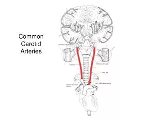
vertebrobasilar system • two vertebral arteries (carrying about 1/3rd of cerebral blood) arise from first part of the subclavian arteries • ascend through foramen transversarium of cervical vertebrae then enter skull through foramen magnum
vertebrobasilar system • major branches of vertebral artery • anterior spinal artery • posterior inferior cerebellar artery • bulbar branches • +/- anterior inferior cerebellar artery • both vertebral arteries unite at the pontomedullary junction to form the basilar artery
vertebrobasilar system • basilar artery continues in midline of pons before it bifurcates into its two terminal posterior cerebral arteries at pontomesencephalic junction • gives off: • anterior inferior cerebellar artery • internal auditory artery • pontine arteries • superior cerebellar artery
the circle of Willis • anterior & posterior communicating arteries help form an arterial circle ‘of Willis’ on the ventral aspect of the brain • provides possibility of collateral circulation in event of occlusion in one of the major arteries proximal to the circle
stroke - definition • “sudden neurological deficit of vascular aetiology lasting more than 24 hours” • compared with TIA (transient ischaemic attack) indicates a transient neurological deficit of vascular origin lasting less than 24 hours
stroke - categorized as • cerebral infarction (80%) • signifying ischaemic brain damage due to occlusion of a vessel • cerebral haemorrhage • primary pathology involves vascular rupture & extravasation of blood into surrounding tissue or compartments
artery territory mca occluson aca occlusion stroke syndrome contralateral hemiplegia, hemianaesthesia, homonymous hemianopia, aphasia, inattention, cortical sensory loss hemiparesis chiefly in the leg stroke
artery territory internal carotid artery occlusion pca occlusion stroke syndrome mixture of aca & mca syndromes (may be asymptomatic) homonymous hemianopia, disconnection syndromes, hemianaesthesia, amnesia, midbrain & thalamic syndromes stroke
artery territory vertebrobasilar thrombosis (basilar occlusion) ventral pontine infarction stroke syndrome quadriparesis, bulbar paralysis, impaired gaze, coma quadriparesis, absent horizontal (but retained vertical) gaze, normal conscious state (‘locked in’ syndrome) stroke
artery territory lateral medullary syndrome (vertebral, pica or aica) stroke syndrome ipsilateral ataxia (icp), Horner’s syndrome, dysphagia & dysarthria (CN 9 & 10), vertigo, nausea & Nystagmus (vest. nuclei), ipsilateral facial anaesthesia (CN 5), contralateral pain & temperature loss (spinothalamic tract) stroke
artery territory medial medullary syndrome stroke syndrome ipsilateral paralysis of tongue (CN 12), contralateral hemiparesis (corticospinal tract), contralateral impairment of touch & position sense (medial lemniscus) stroke
venous drainage • superior sagittal sinus --> right transverse sinus (confluence of sinuses) --> right sigmoid sinus • inferior sagittal sinus --> straight sinus --> left transverse sinus (confluence of sinuses) --> left sigmoid sinus • the sigmoid sinuses --> (become) internal jugular veins at jugular foramen
cavernous sinus • located lateral aspect sphenoid • receives blood from pituitary gland, orbit through ophthalmic veins & middle cerebral veins • cavernous sinus --> superior & inferior petrosal sinuses
cavernous sinus • superior petrosal sinus --> junction transverse & sigmoid sinuses • inferior petrosal sinus --> internal jugular vein • emissary veins link CS with facial & scalp veins
cerebrospinal fluid circulation • clear & colorless water-like fluid • formed by choroid plexus • mainly in lateral ventricles (& to lesser degree in 3rd & 4th ventricles) • formation of CSF complex • includes both passive filtration & active secretary mechanisms
CSF circulation • CSF produced in lateral ventricles • enters third ventricle through interventricular foramen • flows through cerebral aqueduct • into fourth ventricle • from fourth ventricle it reaches the subarachnoid space
CSF circulation • CSF enters subarachnoid space via three openings: • median aperture (posterior medullary velum) • two lateral apertures (lateral recesses of fourth ventricle)
CSF circulation • collections of microscopic arachnoid villi form macroscopic elevations (arachnoid granulations) that protrude into the lateral expansions of the superior sagittal sinus through openings in the dura • flow of CSF is fairly rapid
CSF circulation • total volume of CSF in the ventricular system & subarachnoid space is only about 125 ml • but it is estimated that about four times that amount (~500 ml) is formed during a 24 hr period • a small amount of CSF seeps down around the spinal cord