Circulation
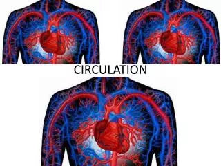
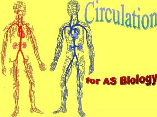
Circulation. for AS Biology. Double circulation. Mammals have a double circulation The right side of the heart sends blood to the lungs ( pulmonary circulation )

Circulation
E N D
Presentation Transcript
Circulation for AS Biology
Double circulation • Mammals have a double circulation • The right side of the heart sends blood to the lungs (pulmonary circulation) • Blood returns from the lungs to the left side of the heart which pumps it into the systemic circulation supplying the rest of the body
Double circulation • What is the advantage of a double circulation?
Double circulation • A double circulation has the advantage of providing all body organs with oxygenated blood at high pressure In the single circulation of a fish, blood is pumped Heart Organs Gills Most of the pressure generated by the heart is lost in the gills, and the organs receive low pressure oxygenated blood.
Double circulation • Double circulation is made possible by the complete division of the heart into separate left and right sides Pulmonary circulation • The left side receives oxygenated blood from the lungs, and pumps it to the systemic circulation; the right side receives deoxygenated blood from the body, and pumps it to the lungs. Systemic circulation
The diagram shows the heart at diastole – relaxed, between contractions. Structure of the heart Semilunar valves Pulmonary artery Pulmonary veins Superior vena cava Aorta Right atrium Left atrium Right atrio-ventricular valve (tricuspid valve) Left atrio-ventricular valve (bicuspid valve) Inferior vena cava Right ventricle Septum Left ventricle Tendon Papillary muscle
The cardiac cycle At ventricular systole (0.3 s): the ventricles contract; ventricular pressure rises above atrial, closing the atrioventicular valves; ventricular pressure rises above arterial, opening the semilunar valves; blood is forced from ventricles into the pulmonary artery and aorta. At atrial systole (0.1 s): the atria contract; the vein openings are constricted, preventing backflow into the veins blood is forced from atria into ventricles through the open atrioventicular valves; the semilunar valves remain closed, because arterial pressure is still higher than ventricular pressure. At diastole (0.4 s): the heart muscle is relaxed; the atrioventicular valves are open, and blood flows passively from atria into ventricles; the semilunar valves are closed, because arterial pressure is higher than ventricular pressure. Papillary muscles adjust tension in valve tendons
Pressure changes during the cardiac cycle This wave of arterial pressure is felt as the pulse This is the arteries’ elastic recoil At ventricular systole the ventricle walls contract and ventricular pressure rises steeply As soon as ventricular pressure rises above atrial, the atrio-ventricular (bicuspid) valve closes, making the ‘lubb’ sound In this sequence we will look at changes in atrial, ventricular and aortic pressure in the left side of the heart during one cardiac cycle. As soon as ventricular pressure falls below atrial, the atrioventricular valve opens and blood flows passively from atria into ventricles In atrial systole, atrial pressure rises as the walls of the atria contract … The aortic semi-lunar valves open as ventricular pressure rises above aortic, then close as it falls below again As blood is forced into the ventricles ventricular pressure rises too, but remains below atrial.
Control of the cardiac cycle • Contraction of cardiac muscle is myogenic: cardiac muscle cells contract of their own accord, without being stimulated by nerves • Isolated cardiac muscle cells will contract in their own independent rhythm • The cardiac cycle is initiated by waves of electrical excitation generated by the sino-atrial node, a specialised patch of muscle in the wall of the right atrium
Control of the cardiac cycle 0.1 s 0.4 s The muscular walls of the atria are insulated from the ventricle walls by a ring of fibrous tissue, the annulus fibrosus: excitation passes to the ventricles by being picked up by the atrioventricular node (AVN) and sent down the Bundle of His, passing to the ventricle walls via the Purkinje fibres. 0.3 s The ventricles contract from the apex upwards, forcing blood upward into the arteries.
Control of the cardiac cycle • Because cardiac muscle contraction is myogenic, if the heart’s nerve supply is severed it will continue to beat • The sino-atrial node is innervated by two nerves, a sympathetic nerve and a branch of the vagus nerve (cranial X), part of the parasympathetic system • Stimulation of the vagus nerve (parasympathetic) slows the heart rate • Sympathetic stimulation speeds the heart rate
Coronary circulation Aorta The muscular wall of the heart is provided with high-pressure oxygenated blood by the coronary arteries, arising from the base of the aorta.
Blood vessels • Arteries carry blood from the heart towards capillary beds, veins carry blood from capillary beds towards the heart • Arteries do not always carry oxygenated blood, nor veins deoxygenated: the pulmonary and umbilical arteries carry deoxygenated blood, the pulmonary and umbilical veins oxygenated blood
Arteries Arteries characteristically have: a narrow lumen, maintaining high pressure a thick wall to withstand high pressure a corrugated inner lining (endothelium), allowing stretching during systole extensive elastic tissue, which absorbs some of the energy given to the blood at systole and then returns it by recoiling during diastole muscle fibres, allowing the artery to be constricted or dilated to control the amount of blood flowing through it (NB this muscle does not propel the blood)
Artery structure - single layer of epithelial cells - on which epithelial cells rest - absorbs energy and recoils - vasoconstriction and vasodilation - absorbs energy and recoils - mostly collagen fibres, holding the artery together
Veins Veins characteristically have: a wide lumen, giving minimum resistance to low pressure flow a smooth endothelium, again giving minimum resistance little elastic or muscular tissue valves to prevent backflow of low pressure blood
Arteries, capillaries and veins As arteries branch and become smaller, their muscle and elastic layers are reduced. Arterioles still have muscle and a nerve supply, and control the blood supply to capillary beds Capillaries have no muscle or nerve supply, only a single cell layer (the endothelium) As capillaries rejoin they form venules, which reunite to form veins
Capillary structure The capillary wall is a single layer of squamous epithelial cells. It is highly permeable, with gaps between cells and fenestrations through cells: capillary beds are where exchange occurs between blood and tissues. The lumen of a capillary is about 5 mm in diameter: red blood cells (diameter 7 mm) pass through in single file, squashed against the capillary wall. Nucleus of squamous epithelial cell Capillary lumen Red blood cell Basement membrane of squamous epithelial cell – allows small molecules through, keeps plasma proteins in
Blood pressure and velocity Only diastolic blood pressure is shown (blue line), i.e. without the rise (pulse) at each ventricular systole Where in the circulatory system does the greatest drop in blood pressure occur? What causes it? The greatest drop in blood pressure occurs in the arterioles. As the arterioles branch and form capillaries, the total cross-sectional area of all the vessels combined is about 1000x greater than that of the arteries, leading to the pressure drop.
Formation of tissue fluid At the arterial end of a capillary bed the blood is still at high pressure (about 300 kPa). Dissolved plasma proteins give the blood a lower solute potential than the fluid in the tissues, but the difference in hydrostatic pressure forces water and solutes out. At the venous end blood is at lower pressure (about 120 kPa). Plasma proteins are retained by the capillary’s basement membrane, so the blood’s solute potential is effectively unchanged. The blood’s water potential is now lower than that of the tissue fluid, and water flows back in. yp= 120 kPa ys= -190 kPa yw= -70 kPa yp= 300 kPa ys= -190 kPa yw= 110 kPa Tissue yw= -25 kPa
Reabsorption of tissue fluid Some of the water in tissue fluid is reabsorbed by osmosis at the venous end of the capillary bed, as explained in the previous slide, but the water potential difference is greater at the arterial end and more fluid leaves the capillaries than re-enters them. The excess is absorbed by capillaries of the lymphatic system, which returns it to the bloodstream at veins in the thorax (where blood pressure is lowest, especially during inspiration). Lymph vessel Lymphatic capillaries Vein Arteriole Venule Artery Tissue Tissue fluid Tissue
The lymphatic system After tissue fluid has entered the lymphatic vessels it is called lymph. The lacteals in the villi of the small intestine are also branches of the lymphatic system: the fat they absorb gives lymph a milky appearance. On its way back to the bloodstream lymph passes through numerous lymph nodes, concentrated masses of lymphocytes and other leucocytes, which filter out micro-organisms and other foreign particles. The spleen, thymus, tonsils and adenoids are also concentrated masses of lymphoid tissue, as are Peyer’s patches in the gut wall.
Practice questions The micrograph shows a blood vessel in transverse section • Identify the type of blood vessel shown and give two reasons for your answer. • (3 marks) • Name two kinds of tissue you would expect to find in the layer labelled X, and state one function for each. • (2 marks) X
Practice questions A B • Identify the stage of the cardiac cycle shown in the diagram. Give two reasons for your answer. • (3 marks) • Identify the blood vessels labelled A, B and C. • (3 marks) • Explain the difference in thickness of the muscular wall at X and Y. • (2 marks) C X Y
Checklist Make sure you • understand the outline functions of the circulatory system in the transport of respiratory gases, metabolites, metabolic wastes and hormones • can describe the double circulatory system • can describe the structure of the mammalian heart and coronary circulation • understand the cardiac cycle; myogenic stimulation • understand how the cardiac cycle is coordinated
Checklist Make sure you • can describe the structure and role arteries, veins and capillaries • can describe the interchange of materials between capillaries and tissue fluid, including the formation and reabsorption of tissue fluid