Circulation
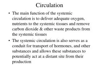
Circulation. Chapter 23. Cardiovascular System. Accepts oxygen, nutrients, and other substances from the respiratory and digestive systems and delivers them to cells Accepts carbon dioxide and wastes from cells and delivers them to respiratory and urinary systems for disposal.

Circulation
E N D
Presentation Transcript
Circulation • Chapter 23
Cardiovascular System • Accepts oxygen, nutrients, and other substances from the respiratory and digestive systems and delivers them to cells • Accepts carbon dioxide and wastes from cells and delivers them to respiratory and urinary systems for disposal
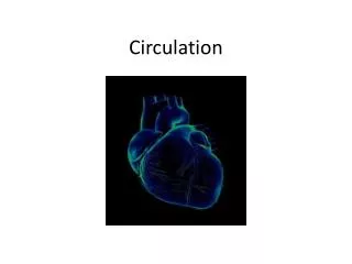
Chambers of heart • RIGHT ATRIUM • RIGHT VENTRICLE • LEFT ATRIUM • LEFT VENTRICLE
CO2 O2 CO2 CO2 Lung Lung O2 O2 Heart O2 O2-rich blood O2-poor blood CO2 (a) Pulmonary circuit (b) Systemic circuit Figure 23.2
One complete trip through the human cardiovascular system: • Takes about one minute • Requires two passes through the heart Animation: Path of Blood in Mammals
Capillaries of head, chest, and arms Superior vena cava Pulmonary artery Pulmonary artery Aorta Capillaries of lung Capillaries of lung Pulmonary vein Pulmonary vein Right atrium Left atrium Right ventricle Left ventricle Inferior vena cava Capillaries of abdominal region and legs O2-rich blood O2-poor blood Figure 23.3-11
To body O2-rich blood From body O2-poor blood Right lung Left lung Right atrium Left atrium Valves Valves Left ventricle Right ventricle From body Figure 23.4
TheCardiac Cycle • The heart relaxes and contracts throughout our lives. • Diastole is the relaxation phase of the heart cycle. • Systole is the contraction phase. Blast Animation: Cardiac Cycle Overview
Atria contract. Blood is forced into ventricles. Heart is relaxed. Blood flows in. 0.1 sec Diastole Systole 0.8 sec 0.3 sec 0.4 sec Ventricles contract. Blood is pumped out. Figure 23.5-3
The Pacemaker and the Control of Heart Rate • The pacemaker, or SA (sinoatrial) node: • Sets the tempo of the heartbeat • Is composed of specialized muscle tissue in the wall of the right atrium Blast Animation: Electrical Coordination of the Cardiac Cycle
Wire leading to SA node Heart Artificial pacemaker Pacemaker (SA node) AV node Right atrium Right ventricle Impulses spread through atria. Pacemaker generates electrical impulses. Impulses reach ventricles. (b) Artificial pacemaker (a) The heart’s natural pacemaker Figure 23.6
Blood Vessels • If the heart is the body’s “pump,” then the “plumbing” is the system of arteries, veins, and capillaries. • Arteries carry blood away from the heart. • Veins carry blood toward the heart. • Capillaries allow for exchange between the bloodstream and tissue cells.
Blood Flow through Arteries • The force that blood exerts against the walls of blood vessels is blood pressure. • Blood pressure is the main force driving the blood from the heart to the capillary beds. • A pulse is the rhythmic stretching of the arteries caused by the pressure of blood forced into the arteries during systole.
From heart To heart Epithelium Valve Epithelium Epithelium Smooth muscle Smooth muscle Connective tissue Connective tissue Artery Vein Venule Arteriole Capillary Figure 23.8
Capillary Tissue cell Red blood cell Diffusion of CO2 and wastes out of tissue cells and into capillary Diffusion of O2 and nutrients out of capillary and into tissue cells From artery To vein Interstitial fluid To vein LM (b) Chemical exchange (a) Capillaries Figure 23.9
Blood Flow through Capillary Beds • At any given time, only about 5–10% of the capillaries have a steady flow of blood. • The regulation of blood flow through capillaries • Is controlled by muscles • Reflects shifting demands by organs of the body
Capillaries • The walls of capillaries are thin and leaky. • At the arterial end of the capillary, blood pressure pushes fluid rich in oxygen, nutrients, and other substances into the interstitial fluid. • At the venous end of the capillary CO2 and other wastes diffuse from tissue cells into the capillary bloodstream.
Blood moves back towards the heart because of: • Surrounding skeletal muscles that compress the veins • One-way valves that permit blood flow only toward the heart
Blood • An adult human has about 5 L (11 pints) of blood. • By volume, blood is about: • 45% cells and • 55% plasma, consisting of about: • 90% water • 10% dissolved salts, proteins, and other molecules
Plasma (55%) Cellular elements (45%) Red blood cells (erythrocytes) Water (90% of plasma) Proteins Blood Dissolved salts (such as sodium, potassium, calcium) White blood cells (leukocytes) Substances being transported (such as O2, CO2, nutrients, wastes, hormones) Platelets Figure 23.11
Blood Cells • Red blood cells (erythrocytes) • Contain hemoglobin • Rapidly transport oxygen (and carbon dioxide) • Have no nucleus when mature • White blood cells (leukocytes) • Tissue maintenance and repair • Defenses against pathogens
Red Blood Cells • ABOUT 99% OF ALL BLOOD CELLS • FUNCTION TO TRANSPORT OXYGEN AND CARBON DIOXIDE • PICK UP IN LUNGS • DELIVER TO ALL CELLS • CONTAIN IRON RICH PROTEIN HEMOGLOBIN • MADE IN BONE MARROW CONTINUOUSLY Fig. 34.6, p. 558
CIRCULATE FOR ABOUT 120 DAYS THEN ARE BROKEN DOWN CAN’T REPLICATE - THEY HAVE NO NUCLEUS ARE MADE IN THE BONE MARROW RBC in capillary Red Blood Cells
Platelets • Platelets function in blood clotting • Platelets and all blood cells arise from stem cells in bone marrow
White Blood Cells • LEUKOCYTES - 1% OF BLOOD CELLS • NORMAL BLOOD -ABOUT 10,000 CELLS/MM3 • MAKE IN THE BONE MARROW • ARE NUCLEATED • HELP BODY DEFEND AGAINST INVADERS • LEUKEMIA CANCER OF WHITE BLOOD CELLS
WBC • 5 KINDS OF WHITE BLOOD CELLS • NEUTROPHILS-PHAGOCYTOSIS AND PROCESSING • LYMPHOCYTES- IMMUNITY • MONOCYTES-GIVE RISE TO MACROPHAGES • EOSINOPILS- ASSOCIATED WITH ALLERGIES • BASOPHILS-INTENSIFY INFLAMMATION
Blood Platelets • FRAGMENTS OF CELLS • FUNCTION IN CONTROLLING BLEEDING • FORMED IN THE BONE MARROW
Platelets (bits of membrane-enclosed cytoplasm that aid clotting) Colorized SEM Fibrin Colorized SEM Red blood cell Figure 23.12c
ABO Blood Typing • Helps match blood of donors and recipients to avoid blood transfusion problems (agglutination)
Rh Blood Typing • Helps prevent problems that may arise when maternal and fetal Rh blood types differ
Circulatory System Disorders • Atherosclerosis, hypertension (chronic high blood pressure), heart attacks, strokes, certain arrhythmias • Regular exercise, maintaining normal body weight, and not smoking lower risk for these disorders
Plaque Connective tissue Smooth muscle Epithelium Normal artery Artery partially blocked by plaque Figure 23.14
Aorta Coronary artery (supplies oxygen to the heart muscle) Dead muscle tissue Blockage Figure 23.13