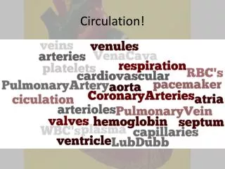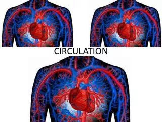
Circulation
Circulation. 2 / 27. Types of circulatory systems. diffusion is slow – circulation (bulk flow) for the distribution of oxygen and nutritients other solutions also exist – trachea in insects, intense enter ic system in parasites, etc. open circulation low pressure, slow flow


Circulation
E N D
Presentation Transcript
2/27 Types of circulatory systems • diffusion is slow – circulation (bulk flow) for the distribution of oxygen and nutritients • other solutions also exist – trachea in insects, intense enteric system in parasites, etc. • open circulation • low pressure, slow flow • slow way of living • exception – insects, because of the tracheal system • closed circulation • high pressure, fast flow, fast regulation • active life • e.g. difference between snails and cephalopods; vertebrates and invertebrates in general • further development: separated circuits in vertebrates for the body and lungs – high pressure would cause filtration in lungs
3/27 Circulatory system of mammals Eckert: Animal Physiology, W.H.Freeman and Co., N.Y.,2000, Fig. 12-3.
4/27 Human heart Berne and Levy, Mosby Year Book Inc, 1993, Fig. 24-10
5/27 Valves in the heart Berne and Levy, Mosby Year Book Inc, 1993, Fig. 24-11
6/27 Electrical activity of the heart • vertebrate heart is miogenic – see Aztec rituals • principal pacemaker: sinoatrial node • 2x8 mm, built up by modified muscle cells • AP is followed by slow hypopolarization – hyperpolarization induced mixed channels (Na+, Ca++) and K+ inactivation • NA and ACh changes the pacemaker potential in different directions through cAMP effecting the hyperpolarization induced channel • in the atrium – rudimentary conduction system • AV-node, 22x10x3 mm, in the interatrial septum • bundle of His, bundle branches (Tawara), Purkinje fibers • SA, AV nodes 0.02-0.1 m/s, muscle cell 0.3-1 m/s, specialized fibers 1-4 m/s (70-80 vs. 10-15 ) Berne and Levy, Mosby Year Book Inc, 1993, Fig. 23-25
7/27 Electrocardiogram • fibers in the atria and in the ventricles are activated in synchrony • high-amplitude signal • vector and scalar ECG • Einthoven triangle • diagnostic importance - infarction, angina • mean electrical axis • arrhythmias • premature depolarization extrasystole, compensatory pause • fibrillation - atrial, ventricular (soap operas) Eckert: Animal Physiology, W.H.Freeman and Co., N.Y.,2000, Fig. 12-8.
8/27 Cardiac cycle Berne and Levy, Mosby Year Book Inc, 1993, Fig. 24-13
9/27 Regulation of cardiac output I. • cardiac output = heart rate x stroke volume • frequency is regulated mainly by the autonomic nervous system • stroke volume depends on the myocardial performance that in turns depends on intrinsic and extrinsic factors • heart rate at rest is about 70/minute • during sleep it is less by 10-20, in children and small animals it can be much higher (hummingbird) • emotional excitation, exercise: 120-150 • parasympathetic inhibition dominates in rest from the vagal nerves – ganglion on the surface or in the wall of the heart • asymmetric: right - SA, left - AV • acting through muscarinic receptors • beat-to-beat regulation – fast elimination
10/27 Regulation of cardiac output II. • sympathetic innervation: lower 1-2 cervical, upper 5-6 dorsal segments • relay in stellate ganglion • beta adrenergic effect through cAMP - positive chronotropic, inotropic, dromotropic, batmotropic effects • slow effect, slow elimination • asymmetric innervation: right - frequency, left – strength of contraction • other effects: • baroceptor reflex • respiratory sinus arrhythmia: rate increases during inspiration, decreases during expiration • explanation: vagal activity increases during expiration (asthma), filling of the heart (preload) increases frequency
11/27 Myocardial performance • intrinsic factors: Starling´s law of the heart, or the Frank-Starling mechanism - 1914 • myocardial performance increases with preload to a certain extent • length of skeletal muscles is optimal at rest, length of heart muscles is optimal when stretched • increased preload: • first the heart cannot pump out the increased venous volume – end systolic volume increases • larger end-diastolic volume – stronger contraction – new equilibrium, increased volume is pumped out • increased peripheral resistance: • first the heart cannot pump out the same volume against the increased resistance – end systolic volume increases • larger end-diastolic volume – stronger contraction – new equilibrium, increased volume is pumped out • extrinsic factors: most important: sympathetic effect – strength of contraction increases Berne and Levy, Mosby Year Book Inc, 1993, Fig. 25-16
12/27 Hemodynamics • circulation cannot be described by simple physical rules: those apply for homogenous fluids flowing by laminar flow in rigid tubes • it is worth to examine those rules as we don’t have any other choice • basic principles: • v - linear velocity (cm/s) • Q - flow (cm3/s) • v = Q / A – linear velocity depends on the cross sectional area • Q = P / R – analogous to Ohm’s law P * * r4 8 * * l Q = ---------- R = --------- 8 * * l * r4 • arterioles have high resistance because of the small „r” • relations between viscosity and hematocrit • turbulent and laminar flow – measurement of blood pressure Eckert: Animal Physiology, W.H.Freeman and Co., N.Y.,2000, Fig. 12-26. Eckert: Animal Physiology, W.H.Freeman and Co., N.Y.,2000, Fig. 12-24.
13/27 The arterial system • large volume, distensible wall, terminated by a large resistance - “Windkessel” • punctured tire, Scotch pipe, etc. • small variation in pressure, continuous flow • decreases the workload of the heart – heart should pump stroke volume in 1/6 time • terms: systolic/diastolic pressure, pulse pressure, mean arterial pressure • mean arterial pressure depends on the blood volume in the arterial system and on the distensibility of the walls of the arteries • pulse pressure depends on stroke volume and compliance • heart copes with increased venous return and peripheral pressure through the arterial system Berne and Levy, Mosby Year Book Inc, 1993, Fig. 27-13 Eckert: Animal Physiology, W.H.Freeman and Co., N.Y.,2000, Fig. 12-28. Berne and Levy, Mosby Year Book Inc, 1993, Fig. 27-10
14/27 Microcirculation I. • in most tissues cells are less than 3-4-cells distance from the nearest capillary • length 1 mm, diameter 3-10 • arteriole - metarteriole - precapillary sphincter - capillary - pericytes • arteriovenous anastomosis (shunt) • nutritional and non-nutritional circulation (thermoregulation) – rat’s tail, rabbit’s ear, etc. • growth of capillaries depends on demand – babies born before term are put into incubators – upon removal, lens are invaded by capillaries - blindness • capillary permeability depends on location (function) • easy penetration for lipid soluble substances • for hydrophilic ones it depends on capillary type Eckert: Animal Physiology, W.H.Freeman and Co., N.Y.,2000, Fig. 12-36.
15/27 Microcirculation II. • continuous capillary • continuous basal membrane, gaps of 4 nm, 7 nm pinocytotic vesicles • muscle, nervous tissue, lung, connective tissue, exocrine glands • fenestrated capillary • continuous basal membrane, pores • everything can penetrate, except proteins and blood cells • kidney, gut, endocrine glands • sinusoidal capillary • large paracellular gaps crossing through the basal membrane • liver, bone marrow, lymph nodes, adrenal cortex • hydrostatic pressure difference - filtration (2% out, 85% back) – exchange of materials • filtration - reabsorption – Starling’s hypothesis • edema: gravidity, tight socks, heart failure, starving, inflammation, elephantiasis, Eckert: Animal Physiology, W.H.Freeman and Co., N.Y.,2000, Fig. 12-36. Eckert: Animal Physiology, W.H.Freeman and Co., N.Y.,2000, Fig. 12-39.
16/27 Regulation of peripheral circulation • central and local regulation – location-, and time-dependent • target: arteriole, metarteriole, sphincter muscles • sympathetic innervation in most cases • strong, long-term contraction without Na-channels – single-unit smooth muscle • local regulation • basal miogenic tone • metabolic regulation - adenosine • external regulation: sympathetic vasoconstriction • parasympathetic effect e.g. on saliva glands, possibly artifact (bradykinin) • humoral effect: NA in low concentration dilates ( -adrenergic), in high concentration contracts (-adrenergic) vessels
17/27 Venous system • veins have thin-walls and large volume – capacity vessels • maximal pressure is about 11 mmHg, but contains half of the blood volume • effect of gravitation: U-shaped tube, pressure difference is the same standing and laying – hydrostatic pressure is huge at the turn • role of the muscle pump and the valves • effect of inspiration – Valsalva's maneuver; in trumpet players - pressure can be around 100-400 mmHg • thrombus and embolus • venomotor tone – standing in attention, fighter pilots, circulatory shock, returning of astronauts • jumping out of bed - 3-800 ml displaced into legs – cardiac output decreases by 2 l
18/27 Coupling of heart and vessels I. • balance of blood flow: pumped volume = volume flowing through the periphery • pumped volume is influenced by: heart beat, contractility of heart, preload, afterload • the last two are coupling factors – influenced by both the heart and the vessels • two important relationships should be considered • cardiac function curve – Starling’s law – vessels can be replaced by tubes • vascular function curves – increase of cardiac output decreases central venous pressure – heart can be replaced by a pump • blood is moved by the difference of mean arterial pressure and central venous pressure • these pressure values depend on blood volume and compliance in the respective systems Berne and Levy, Mosby Year Book Inc, 1993, Fig. 30-6 Berne and Levy, Mosby Year Book Inc, 1993, Fig. 30-7
19/27 Coupling of heart and vessels II. • circulation stops: mean circulatory pressure, when heart restarts it pumps blood from the venous into the arterial system • the two function curves act against each other – moving water in a circular channel • equilibrium is reached – many phenomena, such as heart failure can be explained by this • in case of physical exercise, bleeding, veins contract – internal infusion • sympathetic effect – cardiac function curve is shifted upward • increase in peripheral resistance shifts both curves downward – balance depends on several factors Berne and Levy, Mosby Year Book Inc, 1993, Fig. 30-2 Berne and Levy, Mosby Year Book Inc, 1993, Fig. 30-9 Berne and Levy, Mosby Year Book Inc, 1993, Fig. 30-12
20/27 Coronary circulation I. • heart cannot accumulate oxygen dept, as it never stops – it is always aerobic • at rest consumption is 8-10 ml/100g/min O2, that is 25-30 ml/min for a 300 g male heart • total consumption is 250 ml/min, this is 12% of that • the stopped heart (dog) has a consumption of 2 ml/100g/min O2 • O2 extraction is very high: venous blood has a saturation of about 25% (20 mmHg) • during physical exercise blood flow can only increase from 180-240 ml/min to 900-1200 ml/min • many mitochondria, scarce mioglobin and glycogen • utilizes everything: glucose, lactate, fatty acids, ketone bodies, amino acids
21/27 Coronary circulation II. • coronary arteries – surround the heart • very good blood supply, 8-10 times as many open capillaries than in the functioning skeletal muscle – almost one capillary/muscle cell • hypertrophy can deteriorate nutrition • end-artery system: almost no overlap – fast occlusion, <10% blood, slow – anastomosis • myocardial infarct - necrosis • blood supply of left ventricle stops during systole • heart rate – systole/diastole ratio! • autoregulation is crucial (between 60-180 mmHg), basal miogenic tone, metabolic regulation (adenosine, NO) • NA – indirect vasodilatation, direct (weak) vasoconstriction – coronary constriction, emotional excitation, increased O2 need – death can occur
22/27 Cerebral circulation • high O2 (3-4 ml/100g/min) and glucose need • irreversible damage after 3-6 minutes • mass is 2% of bodyweight, circulation 15%, O2 consumption 25% • circle of Willis – right and left a. carotis and a. vertebralis – moderate contralateral circulation • specialty: closed space – constant blood flow • local differences, measurement: 85Kr, 133Xe, PET • tumor, bleeding, edema – severe consequences • autoregulation very strong between 60-160 mmHg - mechanism unknown • metabolic regulation: adenosine, NO, CO2, K+ • no sympathetic tone at rest • 3 compartments: intracellular, interstitial, liquor • interstitial fluid and liquor: low protein content, compared to plasma lower K+ , higher NaCl • blood-brain, blood-liquor barrier, circumventricular windows
23/27 Splanchnic circulation • gastrointestinal tract, liver, spleen, pancreas • blood flow 25 % of total, strongly variable • 18 % of total blood volume (1 l) – blood store, half can be released into circulation • the liver (1.5 kg) uses 20% of total O2 consumption • a. hepatica, hepatic portal vein - portal circulation – effectiveness of pills and suppositories • some local regulation (functional hyperemia after meals), increase is only 50 % • strong central regulation by the sympathetic nervous system • splanchnic vasoconstriction compensates for vasodilatation in muscles during exercise, peripheral resistance and blood pressure stable • constriction of veins provides internal transfusion • in some animals the spleen is a blood store
40-50% of body mass (young man), 20 % of circulation strong exercise: circulation 20 l, muscles get 80% at rest: central regulation, during exercise: metabolic - K+, adenosine (?), H+ on blood vessels not only 1 adrenoreceptors, but 2 as well – more sensitive for adrenalin - dilatation main effect still constriction stimulation of the limbic system, hypothalamus, cortex – in some species dilatation - cholinergic sympathetic fibers role: preparation for exercise, simulated death redistribution of blood flow during exercise – overall vasoconstriction (resistance), venous contraction (volume) – percentages change, but increase of cardiac output more important 24/27 Skeletal muscle circulation
25/27 Skin circulation • typical non-nutritional blood flow • 100 ml/min would be sufficient, but 3-500 ml/min flows • serving thermoregulation: thermal exchange and evaporation • at apical parts (fingers, nose, ears, etc.): surface-to-volume ratio large - arteriovenous anastomosis toward the venous plexus – if open, intense flow, more heat loss • strong central regulation: sympathetic innervation, 1 receptor - constriction • dilatation is achieved by a decrease in this tone • sweat glands receive sympathetic cholinergic innervation - bradykinin - dilatation • psychical reactions on the head, neck, upper part of the chest: embarrassment, anger - flushing; fear, anxiety, sorrow - paling; military test
26/27 Central regulation I. rostro-ventro-lateral neurons • regulator neurons are in the medulla (formerly: pressor and depressor centers) – that is why any increase in brain volume can be fatal • input: reflex zones, direct CO2,H+ effect • output: vagal nerve and the sympathetic nervous system – tonic activity at rest: slow heart beat, vasoconstriction in muscle, skin, intestines • chemo-, and mechanoreceptors – information for the control of breathing and for the long-term regulation • part of the receptors found in compact zones, they induce circumscribed reflexes • receptors in the high-pressure system (baroceptors): carotid and aortic sinuses – „buffer nerves” carry the information to the n. tractus solitarius (belongs to the caudal cell group) caudal neurons preganglionic vagal neurons primary afferents sympathetic preganglionic neurons reflexogenic zones sympathetic postganglionic neurons adrenal medulla heart vessels Fonyo, Medicina, 1997, Fig. 23-2
27/27 Central regulation II. • receptors of the low-pressure system (atrial volume receptors): at the orifice of the v. cavae and the v. pulmonalis, as well as at the tip of the ventricles • activated by volume increase, effect similar to baroceptor effect, but long-term responses more important – production of ADH and aldosterone decreases • special receptor group in the atrium: Bainbridge - reflex – filling increase, frequency increase – one factor in sinus arrhythmia • chemoreceptors: glomus caroticum and aorticum activated by CO2 increase and O2 decrease (below 60 mmHg) – latter is more important as CO2 acts also directly in the medulla – heart frequency decreases, vasoconstriction • „sleeping pill” for native people (and biology students): pressing the sinus caroticum
Conduction system of the heart Berne and Levy, Mosby Year Book Inc, 1993, Fig. 23-25
Heart-lung preparation Berne and Levy, Mosby Year Book Inc, 1993, Fig. 25-16
Cross sectional area and velocity Eckert: Animal Physiology, W.H.Freeman and Co., N.Y.,2000, Fig. 12-24.
Pressure changes Eckert: Animal Physiology, W.H.Freeman and Co., N.Y.,2000, Fig. 12-26.
Windkessel function Eckert: Animal Physiology, W.H.Freeman and Co., N.Y.,2000, Fig. 12-28.
Effect of increased venous return Berne and Levy, Mosby Year Book Inc, 1993, Fig. 27-10
Effect of increased resistance Berne and Levy, Mosby Year Book Inc, 1993, Fig. 27-13
Microcirculation Eckert: Animal Physiology, W.H.Freeman and Co., N.Y.,2000, Fig. 12-36.
Types of capillaries Eckert: Animal Physiology, W.H.Freeman and Co., N.Y.,2000, Fig. 12-36.
Starling’s hypothesis Eckert: Animal Physiology, W.H.Freeman and Co., N.Y.,2000, Fig. 12-39.
Effect of blood volume changes Berne and Levy, Mosby Year Book Inc, 1993, Fig. 30-6
Effect of resistance changes Berne and Levy, Mosby Year Book Inc, 1993, Fig. 30-7
Model of the circulatory system Berne and Levy, Mosby Year Book Inc, 1993, Fig. 30-2
Coupling between the heart and the vasculature Berne and Levy, Mosby Year Book Inc, 1993, Fig. 30-12
Effect of sympathetic tone Berne and Levy, Mosby Year Book Inc, 1993, Fig. 30-9
Autonomic nervous system Eckert: Animal Physiology, W.H.Freeman and Co., N.Y.,2000, Fig. 11-15.
Spinal nerves Kiss-Szentagothai, Medicina, 1964, Fig. III-90
Regulation of circulation rostro-ventro-lateral neurons caudal neurons preganglionic vagal neurons primary afferents sympathetic preganglionic neurons reflexogenic zones sympathetic postganglionic neurons adrenal medulla heart vessels Fonyo, Medicina, 1997, Fig. 23-2
Carotid sinus Berne and Levy, Mosby Year Book Inc, 1993, Fig. 29-8
Autonomic nervous system I. • peripheral and central nervous system - PNS-CNS • CNS: brain + spinal chord • PNS: nerves + ganglia (sensory and vegetative) + (enteric nervous system) • afferent and efferent, somatic and vegetative • afferent: similar organization – primary afferent neuron outside of the CNS • efferent: location of motoneurons, outflow, peripheral relaying, transmitter, target • there are two divisions of the autonomic nervous system: sympathetic and parasympathetic • vegetative fibers in spinal nerves • not every organ is innervated by both divisions, effect is not everywhere antagonistic • sympathetic: general effects • parasympathetic: localized effects
Autonomic nervous system II. • preganglionic fibers (B-fibers) • sympathetic and parasympathetic: ACh binding to neuronal nicotinic ACh receptors - ionotropic, opening of Na+-K+ mixed channel - antagonist: e.g. hexamethonium • postganglionic fibers (C-fibers) • parasympathetic: ACh binding to muscarinic ACh receptors - IP3 increase, and opening (direct effect) or closing (indirect effect) of K+-channels - antagonist: atropine, agonist: carbachol • sympathetic: 90% NE, 10% ACh (salivary gland, sweat gland, vasodilatation in skeletal muscles) • 1 - IP3 increase - Ca++ release from internal stores – contraction in smooth muscles, e.g. vessels, sphincters in the intestines, iris radial muscle - NE > Adr • 2 - cAMP decrease – mostly autoreceptor • 1 - cAMP increase - Ca++ - in the heart - chrono-, and inotropic effect - NE = Adr • 2 - cAMP increase - Ca++ pump - relaxation in smooth muscles, e.g. bronchioles, skeletal muscle vessels - Adr >> NE • - agonist: phenylephrine, antagonist: phenoxybenzamine • - agonist: isoprotenerol, antagonist: propanolol