CIRCULATION
850 likes | 1.34k Vues
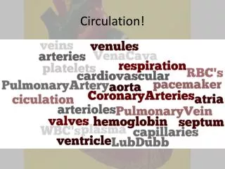
CIRCULATION. Every organism must exchange materials and energy with its environment, and this exchange ultimately occurs at the cellular level. Cells live in aqueous environments. The resources that they need, such as nutrients and oxygen, move across the plasma membrane to the cytoplasm.

CIRCULATION
E N D
Presentation Transcript
Every organism must exchange materials and energy with its environment, and this exchange ultimately occurs at the cellular level. Cells live in aqueous environments. The resources that they need, such as nutrients and oxygen, move across the plasma membrane to the cytoplasm. Metabolic wastes, such as carbon dioxide, move out of the cell. Introduction
Most animals have organ systems specialized for exchanging materials with the environment, and many have an internal transport system that conveys fluid (blood or interstitial fluid) throughout the body. For aquatic organisms, structures like gills present an expansive surface area to the outside environment. Oxygen dissolved in the surrounding water diffuses across the thin epithelium covering the gills and into a network of tiny blood vessels (capillaries). At the same time, carbon dioxide diffuses out into the water.
Diffusion alone is not adequate for transporting substances over long distances in animals - for example, for moving glucose from the digestive tract and oxygen from the lungs to the brain of mammal. 1. Transport systems functionally connect the organs of exchange with the body cells: an overview
Diffusion is insufficient over distances of more than a few millimeters, because the time it takes for a substance to diffuse to one place to another is proportional to the square of the distance. For example, if it takes 1 second for a given quantity of glucose to diffuse 100 microns, it will take 100 seconds for it to diffuse 1 mm and almost three hours to diffuse 1 cm. The circulatory system solves this problem by ensuring that no substance must diffuse very far to enter or leave a cell.
The bulk transport of fluids throughout the body functionally connects the aqueous environment of the body cells to the organs that exchange gases, absorb nutrients, and dispose of wastes. For example, in the mammalian lung, oxygen from inhaled air diffuses across a thin epithelium and into the blood, while carbon dioxide diffuses out. Bulk fluid movement in the circulatory system, powered by the heart, quickly carries the oxygen-rich blood to all parts of the body. As the blood streams through the tissues within microscopic vessels called capillaries, chemicals are transported between blood and the interstitial fluid that bathes the cells.
The body plan of a hydra and other cnidarians makes a circulatory system unnecessary. A body wall only two cells thick encloses a central gastrovascular cavity that serves for both digestion and for diffusion of substances throughout the body. The fluid inside the cavity is continuous with the water outside through a single opening, the mouth. Thus, both the inner and outer tissue layers are bathed in fluid. 2. Most invertebrates have a gastrovascular cavity or a circulatory system for internal transport
In cnidarians like Aurelia, the mouth leads to an elaborate gastrovascular cavity that has branches radiating to and from the circular canal. The products of digestion in the gastrovascular cavity are directly available to the cells of the inner layer, and it is only a short distance to diffuse to the cells of the outer layer. Fig. 42.1
Planarians and most other flatworms also have gastrovascular cavities that exchange materials with the environment through a single opening. The flat shape of the body and the branching of the gastrovascular cavity throughout the animal ensure that are cells are bathed by a suitable medium and diffusion distances are short.
For animals with many cell layers, gastrovascular cavities are insufficient for internal distances because the diffusion transports are too great. In more complex animals, two types of circulatory systems that overcome the limitations of diffusion have evolved: open circulatory systems and closed circulatory systems. Both have a circulatory fluid (blood), a set of tubes (blood vessels), and a muscular pump (the heart). The heart powers circulation by using metabolic power to elevate the hydrostatic pressure of the blood (blood pressure), which then flows down a pressure gradient through its circuit back to the heart.
In insects, other arthropods, and most mollusks, blood bathes organs directly in an open circulatory system. There is no distinction between blood and interstitial fluid, collectively called hemolymph. One or more hearts pump the hemolymph into interconnected sinusessurrounding the organs, allowing exchange between hemolymph and body cells. Fig. 42.2a
In insects and other arthropods, the heart is an elongated dorsal tube. When the heart contracts, it pumps hemolymph through vessels out into sinuses. When the heart relaxes, it draws hemolymph into the circulatory through pores called ostia. Body movements that squeeze the sinuses help circulate the hemolymph.
In a closed circulatory system, as found in earthworms, squid, octopuses, and vertebrates, blood is confined to vessels and is distinct from the interstitial fluid. One or more hearts pump blood into large vessels that branch into smaller ones cursing through organs. Materials are exchanged by diffusion between the blood and the interstitial fluid bathing the cells. Fig. 42.2b
The closed circulatory system of humans and other vertebrates is often called the cardiovascular system. The heart consists of one atrium or two atria, the chambers that receive blood returning to the heart, and one or two ventricles, the chambers that pump blood out of the heart. 3. Vertebrate phylogeny is reflected in adaptations of the cardiovascular system
Arteries, veins, and capillaries are the three main kinds of blood vessels. Arteries carry blood away from the heart to organs. Within organs, arteries branch into arterioles, small vessels that convey blood to capillaries. Capillaries with very thin, porous walls form networks, called capillary beds, that infiltrate each tissue. Chemicals, including dissolved gases, are exchanged across the thin walls of the capillaries between the blood and interstitial fluid. At their “downstream” end, capillaries converge into venules, and venules converge into veins, which return blood to the heart.
Arteries and veins are distinguished by the direction in which they carry blood, not by the characteristics of the blood they carry. All arteries carry blood from the heart toward capillaries. Veins return blood to the heart from capillaries.
Metabolic rate is an important factor in the evolution of cardiovascular systems. In general, animals with high metabolic rates have more complex circulatory systems and more powerful hearts than animals with low metabolic rates. Similarly, the complexity and number of blood vessels in a particular organ are correlated with that organ’s metabolic requirements. Perhaps the most fundamental differences in cardiovascular adaptations are associated with gill breathing in aquatic vertebrates compared with lung breathing in terrestrial vertebrates.
A fish heart has two main chambers, one atrium and one ventricle. Blood is pumped from the ventricle to the gills (the gill circulation) where it picks upoxygen and disposes ofcarbon dioxide across thecapillary walls. The gill capillaries convergeinto a vessel that carriesoxygenated blood to capillarybeds at the other organs(the systemic circulation)and back to the heart. Fig. 42.3a
In fish, blood must pass through two capillary beds, the gill capillaries and systemic capillaries. When blood flows through a capillary bed, blood pressure - the motive force for circulation - drops substantially. Therefore, oxygen-rich blood leaving the gills flows to the systemic circulation quite slowly (although the process is aided by body movements during swimming). This constrains the delivery of oxygen to body tissues, and hence the maximum aerobic metabolic rate of fishes.
Frogs and other amphibians have a three-chambered heart with two atria and one ventricle. The ventricle pumpsblood into a forkedartery that splits theventricle’s output intothe pulmocutaneousand systemiccirculations. Fig. 42.3b
The pulmocutaneous circulation leads to capillaries in the gas-exchange organs (the lungs and skin of a frog), where the blood picks up O2 and releases CO2 before returning to the heart’s left atrium. Most of the returning blood is pumped into the systemic circulation, which supplies all body organs and then returns oxygen-poor blood to the right atrium via the veins. This scheme, called double circulation, provides a vigorous flow of blood to the brain, muscles, and other organs because the blood is pumped a second time after it loses pressure in the capillary beds of the lung or skin.
In the ventricle of the frog, some oxygen-rich blood from the lungs mixes with oxygen-poor blood that has returned from the rest of the body. However, a ridge within the ventricle diverts most of the oxygen-rich blood from the left atrium into the systemic circuit and most of the oxygen-poor blood from the right atrium into the pulmocutaneous circuit.
Reptiles also have double circulation with pulmonary (lung) and systemic circuits. However, there is even less mixing of oxygen-rich and oxygen-poor blood than in amphibians. Although the reptilian heart is three-chambered, the ventricle is partially divided.
In crocodilians, birds, and mammals, the ventricle is completely divided into separate right and left chambers. In this arrangement, the left sideof the heart receives and pumpsonly oxygen-rich blood, whilethe right side handles onlyoxygen-poor blood. Double circulation restores pressure to the systemic circuit and prevents mixing of oxygen-rich and oxygen-poor blood. Fig. 42.3c
The evolution of a powerful four-chambered heart was an essential adaptation in support of the endothermic way of life characteristic of birds and mammals. Endotherms use about ten times as much energy as ectotherms of the same size. Therefore, the endotherm circulatory system needs to deliver about ten times as much fuel and O2 to their tissues and remove ten times as much wastes and CO2. Birds and mammals evolved from different reptilian ancestors, and their powerful four-chambered hearts evolved independently - an example of convergent evolution.
In the mammalian cardiovascular system, the pulmonary and system circuits operate simultaneously. The two ventricles pump almost in unison While some blood is traveling in the pulmonary circuit, the rest of the blood is flowing in the systemic circuit. 4. Double circulation in mammals depends on the anatomy and pumping cycle of the heart
To trace the double circulation pattern of the mammalian cardiovascular system, we’ll start with the pulmonary (lung) circuit. Fig. 42.4
The pulmonary circuit carries blood from the heart to the lungs and back again. (1) The right ventricle pumps blood to the lungs via (2) the pulmonary arteries. As blood flows through (3) capillary beds in the right and left lungs, it loads O2 and unloads CO2. Oxygen-rich blood returns from the lungs via the pulmonary veins to (4) the left atrium of the heart. Next, the oxygen-rich blood blows to (5) the left ventricle, as the ventricle opens and the atrium contracts.
The left ventricle pumps oxygen-rich blood out to the body tissues through the systemic circulation. Blood leaves the left ventricle via (6) the aorta, which conveys blood to arteries leading throughout the body. The first branches from the aorta are the coronary arteries, which supply blood to the heart muscle. The next branches lead to capillary beds (7) in the head and arms. The aorta continues in a posterior direction, supplying oxygen-rich blood to arteries leading to (8) arterioles and capillary beds in the abdominal organs and legs. Within the capillaries, blood gives up much of its O2 and picks up CO2 produced by cellular respiration.
Venous return to the right side of the heart begins as capillaries rejoin to form venules and then veins. Oxygen-poor blood from the head, neck, and forelimbs is channeled into a large vein called (9) the anterior (or superior) vena cava. Another large vein called the (10) posterior (or inferior) vena cava drains blood from the trunk and hind limbs. The two venae cavae empty their blood into (11) the right atrium, from which the oxygen-poor blood flows into the right ventricle.
The mammalian heart is located beneath the breastbone (sternum) and consists mostly of cardiac muscle. The two atria have relatively thin walls and function as collection chambers for blood returning to the heart. The ventricles have thicker walls and contract much more strongly than the atria.
A cardiac cycle is one complete sequence of pumping, as the heart contracts, and filling, as it relaxes and its chambers fill with blood. The contraction phase is called systole, and the relaxation phase is called diastole.
For a human at rest with a pulse of about 75 beat per minute, one complete cardiac cycle takes about 0.8 sec. (1) During the relaxation phase (atria and ventricles in diastole) lasting about 0.4 sec, blood returning from the large veins flows into atria and ventricles. (2) A brief period (about 0.1 sec) of atrial systole forces all the remaining blood out of the atria and into the ventricles. (3) During the remaining 0.3 sec of the cycle, ventricular systole pumps blood into the large arteries.
Cardiac output depends on two factors: the rate of contraction or heart rate (number of beats per second) and stroke volume, the amount of blood pumped by the left ventricle in each contraction. The average stroke volume for a human is about 75 mL. The typical resting cardiac output, about 5.25 L / min, is about equivalent to the total volume of blood in the human body. Cardiac output can increase about fivefold during heavy exercise. Heart rate can be measured indirectly by measuring your pulse - the rhythmic stretching of arteries caused by the pressure of blood pumped by the ventricles.
Four valves in the heart, each consisting of flaps of connective tissue, prevent backflow and keep blood moving in the correct direction. Between each atrium and ventricle is an atrioventricular (AV) valve which keeps blood from flowing back into the atria when the ventricles contract. Two sets of semilunar valves, one between the left ventricle and the aorta and the other between the right ventricle and the pulmonary artery, prevent backflow from these vessels into the ventricles while they are relaxing.
The heart sounds we can hear with a stethoscope are caused by the closing of the valves. The sound pattern is “lub-dup, lub-dup, lub-dup.” The first heart sound (“lub”) is created by the recoil of blood against the closed AV valves. The second sound (“dup”) is the recoil of blood against the semilunar valves.
A defect in one or more of the valves causes a heart murmur, which may be detectable as a hissing sound when a stream of blood squirts backward through a valve. Some people are born with heart murmurs. Others are due damage to the valves by infection. Most heart murmurs do not reduce the efficiency of blood flow enough to warrant surgery.
Because the timely delivery of oxygen to the body’s organs is critical for survival, several mechanisms have evolved that assure the continuity and control of heartbeat. Certain cells of vertebrate cardiac muscle are self-excitable, meaning they contract without any signal from the nervous system. Each cell has its own intrinsic contraction rhythm. However, these cells are synchronized by the sinoatrial (SA) node, or pacemaker, which sets the rate and timing at which all cardiac muscle cells contract. The SA node is located in the wall of the right atrium.
The cardiac cycle is regulated by electrical impulses that radiate throughout the heart. Cardiac muscle cells are electrically coupled by intercalated disks between adjacent cells. Fig. 42.7
(1) The SA node generates electrical impulses, much like those produced by nerves that spread rapidly (2) through the wall of the atria, making them contract in unison. The impulse from the SA node is delayed by about 0.1 sec at the atrioventricular (AV) node, the relay point to the ventricle, allowing the atria to empty completely before the ventricles contract. (3) Specialized muscle fibers called bundle branches and Purkinje fibers conduct the signals to the apex of the heart and (4) throughout the ventricular walls. This stimulates the ventricles to contract from the apex toward the atria, driving blood into the large arteries.
The impulses generated during the heart cycle produce electrical currents that are conducted through body fluids to the skin. Here, the currents can be detected by electrodes and recorded as an electrocardiogram (ECG or EKG).
While the SA node sets the tempo for the entire heat, it is influenced by a variety of physiological cues. Two sets of nerves affect heart rate with one set speeding up the pacemaker and the other set slowing it down. Heart rate is a compromise regulated by the opposing actions of these two sets of nerves. The pacemaker is also influenced by hormones. For example, epinephrine from the adrenal glands increases heart rate. The rate of impulse generation by the pacemaker increases in response to increases in body temperature and with exercise.
All blood vessels are built of similar tissues. The walls of both arteries and veins have three similar layers. On the outside, a layer of connective tissue with elastic fibers allows the vessel to stretch and recoil. A middle layer has smooth muscle and more elastic fibers. Lining the lumen of all blood vessels, including capillaries, is an endothelium, a single layer of flattened cells that minimizes resistance to blood flow. 5. Structural differences of arteries, veins, and capillaries correlate with their different functions
Structural differences correlate with the different functions of arteries, veins, and capillaries. Capillaries lack the two outer layers and their very thin walls consist of only endothelium and its basement membrane, thus enhancing exchange. Fig. 42.8
Arteries have thicker middle and outer layers than veins. The thicker walls of arteries provide strength to accommodate blood pumped rapidly and at high pressure by the heart. Their elasticity (elastic recoil) helps maintain blood pressure even when the heart relaxes.
The thinner-walled veins convey blood back to the heart at low velocity and pressure. Blood flows mostly as a result of skeletal muscle contractions when we move that squeeze blood in veins. Within larger veins, flaps of tissues act as one-way valves that allow blood to flow only toward the heart. Fig. 42.9
At any given time, only about 5-10% of the body’s capillaries have blood flowing through them. Capillaries in the brain, heart, kidneys, and liver are usually filled to capacity, but in many other sites, the blood supply varies over times as blood is diverted. For example, after a meal blood supply to the digestive tract increases. During strenuous exercise, blood is diverted from the digestive tract and supplied to skeletal muscles. 6. Transfer of substances between the blood and the interstitial fluid occurs across the thin walls of capillaries
Two mechanisms, both dependent on smooth muscles controlled by nerve signals and hormones, regulate the distribution of blood in capillary beds. In one mechanism, contraction of the smooth muscle layer in the wall of an arteriole constricts the vessel, decreasing blood flow through it to a capillary bed. When the muscle layer relaxes, the arteriole dilates, allowing blood to enter the capillaries.