Circulation
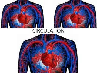
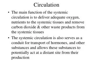
Circulation. The main function of the systemic circulation is to deliver adequate oxygen, nutrients to the systemic tissues and remove carbon dioxide & other waste products from the systemic tissues

Circulation
E N D
Presentation Transcript
Circulation • The main function of the systemic circulation is to deliver adequate oxygen, nutrients to the systemic tissues and remove carbon dioxide & other waste products from the systemic tissues • The systemic circulation is also serves as a conduit for transport of hormones, and other substances and allows these substances to potentially act at a distant site from their production
Functional Parts • systemic arteries • designed to carry blood under high pressure out to the tissue beds • arterioles & pre capillary sphincters • act as control valves to regulate local flow • capillaries- one cell layer thick • exchange between tissue (cells) & blood • venules • collect blood from capillaries • systemic veins • return blood to heart/dynamic storage
Basic theory of circulatory function • Blood flow is proportional to metabolic demand • Cardiac output controlled by local tissue flow • Arterial pressure control is independent of local flow or cardiac output
Characteristics of Vessels • Components • Endothelium- one layer exists in all vessels • Elastic tissue (1) • Smooth muscle (2) • Fibrous tissue (3) • Relative composition • Aorta 1>3>2 • typical artery 2>1>3 • vein 1 = 2 = 3 • capillary- only endothelium
Hemodynamics • Flow • Pressure gradient • Resistance • Ohm’s Law • V = IR (Analogous to P = QR)
Flow (Q) • The volume of blood that passes a certain point per unit time (eg. ml/min) • Q = velocity X cross sectional area • At a given flow, the velocity is inversely proportional to the total cross sectional area • Q = P / R • Flow is directly proportional to P and inversely proportional to resistance (R)
Pressure gradient • Driving force of blood • difference in pressure between two points • proportional to flow (Q) • At a given Q the greater the drop in P in a segment or compartment the greater the resistance to flow.
Resistance • R= 8l/ r4 • = viscosity, l = length of vessel, r = radius • Parallel circuit • 1/RT= 1/R1+ 1/R2 + 1/R3 + … 1/RN • RT < smallest individual R • Series circuit • RT = R1 + R2 + R3 + … RN • RT = sum of individual R’s • The systemic circulation is predominantly a parallel circuit
Advantages of Parallel Circuitry • Independence of local flow control • increase/decrease flow to tissues independently • Minimizes total peripheral resistance (TPR) • Oxygen rich blood supply to every tissue
Viscosity • Internal friction of a fluid associated with the intermolecular attraction • Blood is a suspension with a viscosity of 3 • most of viscosity due to RBC’s • Plasma has a viscosity of 1.5 • Water is the standard with a viscosity of 1 • With blood, viscosity 1/ velocity
Viscosity considerations at microcirculation • velocity decreases which increases viscosity • due to elements in blood sticking together • cells can get stuck at constriction points momentarily which increases apparent viscosity • fibrinogen increases flexibility of RBC’s • in small vessels cells line up which decreases viscosity and offsets the above to some degree (Fahaeus-Lindquist)
Hematocrit • % of packed cell volume (10 RBC’s) • Normal range 38%-45%
Streamline silent most efficient normal Cross mixing vibrational noise least efficient frequently associated with vessel disease (bruit) Laminar vs. Turbulent Flow
Reynold’s number • Probability statement for turbulent flow • The greater the R#, the greater the probability for turbulence • R# = v D / • v = velocity, D = tube diameter, = density, = viscosity • If R# < 2000 flow is usually laminar • If R# > 3000 flow is usually turbulent
Doppler Ultrasonic Flow-meter • Ultrasound to determine velocity of flow • Doppler frequency shift function of the velocity of flow • RBC’s moving toward transmitter, compress sound waves, frequency of returning waves • Broad vs. narrow frequency bands • Broad band is associated with turbulent flow • narrow band is associated laminar flow
Determination of Flow • Determination of Cardiac Output • Fick principal • Indicator dilution • Determination of vessel flow • Venous occlusion plesthymography • Momentary limb blood flow • Doppler ultrasonic flowmeter • Vascular flow cuffs
Fick Principal • Blood flow to a tissue/organ • 3 port system • Input blood concentration of substance x • Output blood concentration of substance x • Addition/removal of substance x from tissue • Flow = amount of substance per min AV difference • See figure 20-18 (Guyton)
Indicator dilution • Based on conservation of mass • CO = mg dye injected X 60 ---------------------------------------------------- ave conc of dye X duration of curve (sec) in each ml for duration of the curve • See figure 20-19 (Guyton)
Distensibility Vs. Compliance • Distensibility is the ability of a vessel to stretch (distend) • Compliance is the ability of a vessel to stretch and hold volume
Distensibility Vs. Compliance • Distensibility = Vol/ Pressure X Ini. Vol • Compliance = Vol/ Pressure • Compliance = Distensibility X Initial Vol.
Volume-Pressure relationships • A volume pressure • In systemic arteries a small volume is associated with a large pressure • In systemic veins a large volume is associated with a small pressure • Veins are about 8 X more distensible and 24 X more compliant than systemic arteries • Wall tone 1/ compliance & distensibility
Control of Blood Flow (Q) • Local blood flow is regulated in proportion to the metabolic demand in most tissues • Short term control involves vasodilatation vasoconstriction of precapillary resist. vessels • arterioles, metarterioles, pre-capillary sphincters • Long term control involves changes in tissue vascularity • formation or dissolution of vessels • vascular endothelial growth factor & angiogenin
Role of arterioles in control of flow • Arterioles act as an integrator of multiple inputs • Arterioles are richly innervated by SNS vasoconstrictor fibers and have alpha receptors • Arterioles are also effected by local factors (e.g.)vasodilators, circulating substances
Local Control of Flow (short term) • Involves vasoconstriction/vasodilatation of precapillary resistance vessels • Local vasodilator theory • Active tissue release local vasodilator (metabolites) which relax vascular smooth muscle • Oxygen demand theory (older theory) • As tissue uses up oxygen, vascular smooth muscle cannot maintain constriction
Local Vasodilators • Adenosine • carbon dioxide • adenosine phosphate compounds • histamine • potassium ions • hydrogen ions • PGE & PGI series prostaglandins
Autoregulation • The ability to keep blood flow (Q) constant in the face of a changing arterial BP • Most tissues show some degree of autoregulation • Q metabolic demand • In the kidney both renal Q and glomerular filtration rate (GFR) are autoregulated
Control of Flow (long term) • Changes in tissue vascularity • On going day to day reconstruction of the vascular system • Angiogenesis-production of new microvessels • arteriogenesis • shear stress caused by enhanced blood flow velocity associated with partial occlusion • Angiogenic factors • small peptides-stimulate growth of new vessels • VEGF (vascular endothelial growth factor)
Changes in tissue vascularity • Stress activated endothelium up-regulates expression of monocyte chemoattractant protein-1 (MCP-1) • attraction of monocytes that invade arterioles • other adhesion molecules & growth factors participate with MCP-1 in an inflammatory reaction and cell death in potential collateral vessels followed by remodeling & development of new & enlarged collateral arteries & arterioles
Changes in tissue vacularity (cont.) • Hypoxia causes release of VEGF • enhanced production of VEGF partly mediated by adenosine in response to hypoxia • VEGF stimulates capillary proliferation and may also be involved in development of collateral arterial vessels • NPY from SNS is angiogenic • hyperactive SNS may compromise collateral blood flow by vasoconstriction • Cancer and angiogenesis
Vasoactive Role of Endothelium • Release prostacyclin (PGI2) • inhibits platelet aggregation • relaxes vascular smooth muscle • Releases nitric oxide (NO) which relaxes vascular smooth muscle • NO release stimulated by: • shear stress associated with increased flow • acetylcholine binding to endothelium • Releases endothelin & endothelial derived contracting factor • constricts vascular smooth muscle
Microcirculation • Capillary is the functional unit of the circulation • bulk of exchange takes place here • Vasomotion-intermittent contraction of metarterioles and precapillary sphincters • functional Vs. non functional flow • Mechanisms of exchange • diffusion • ultrafiltration • vesicular transport
Oxygen uptake/utilization • = the product of flow (Q) times the arterial-venous oxygen difference • O2 uptake = (Q) (A-V O2 difference) • Q=300 ml/min • AO2= .2 ml O2/ml blood • VO2= .15 ml O2/ml blood • 15 ml O2/ min = (300 ml/min) (.05 mlO2/ml)
Functional Vs. Non Functional Flow • Functional or Nutritive flow (Q) is associated with increased oxygen uptake/utilization • x causes Q to from 300 to 600 ml/min, A-V O2 stays at .05 ml O2/ml, O2 uptake has from 15 to 30 ml O2/min, in Q is functional (nutritive) because of O2 uptake • y causes Q to from 300 to 600 ml/min, but A-V O2 from .05 to .025 ml O2/ml, O2 uptake, is still 15 ml O2/min in Q is nonfunctional (non nutritive) because O2 uptake has not changed. • Non nutritive flow increases is associated with shunting of blood through a bed
Capillary Exchange • Passive Diffusion • permeability • concentration gradient • Ultrafiltration • Bulk flow through a filter (capillary wall) • Starling Forces • Hydrostatic P • Colloid Osmotic P • Vesicular Transport • larger MW non lipid soluble substances
Ultrafiltration • Hydrostatic P gradient (high to low) favors filtration • Capillary HP averages 17 mmHg • Interstitial HP averages -3 mmHg • Colloid Osmotic P (low to high) favors reabsorption • Capillary COP averages 28 mmHg • Interstitial COP averages 9 mmHg • Net Filtration P = (CHP-IHP)-(CCOP-ICOP) • 1 = 20 - 19
Colloid Osmotic Considerations • The colloid osmotic pressure is a function of the protein concentration • Plasma Proteins • Albumin (75%) • Globulins (25%) • Fibrinogen (<1%) • Calculated Colloid Effect is 19 mmHg • Actual Colloid Effect is 28 mmHg • Discrepancy is due to the Donnan Effect
Donnan Effect • Increases the colloid osmotic effect • Large MW plasma proteins (1o albumen) carries negative charges which attract + ions (1o Na+) increasing the osmotic effect by about 50%
Effect of Ultrastructure of Capillary Wall on Colloid Osmotic Pressure • Capillary wall can range from tight junctions (e.g. blood brain barrier) to discontinuous (e.g. liver capillaries) • Glomerular Capillaries in kidney have filtration slits (fenestrations) • Only that protein that cannot cross capillary wall can exert osmotic pressure
Reflection Coefficient • Reflection Coefficient expresses how readily protein can cross capillary wall • ranges between 0 and 1 • If RC = 0 • All colloid proteins freely cross wall, none are reflected, no colloid effect • If RC = 1 • All colloid proteins are reflected, none cross capillary wall, full colloid effect
Lymphatic system • Lymph capillaries drain excess fluid from interstitial spaces • No true lymphatic vessels found in superficial portions of skin, CNS, endomysium of muscle, & bones • Thoracic duct drains lower body & left side of head, left arm, part of chest • Right lymph duct drains right side of head, neck, right arm and part of chest
CNS-modified lymphatic function • No true lymphatic vessels in CNS • Perivascular spaces contain CSF & communicate with subarachnoid space • Plasma filtrate & escaped substances in perivascular spaces returned to the vascular system in the CSF via the arachnoid villi which empties into dural venous sinuses • Acts a functional lymphatic system in CNS
Formation of Lymph • Excess plasma filtrate-resembles ISF from tissue it drains • [Protein] 3-5 gm/dl in thoracic duct • liver 6 gm/dl • intestines 3-4 gm/dl • most tissues ISF 2 gm/dl • 2/3 of all lymph from liver & intestines • Any factor that filtration and/or reabsorption will lymph formation
Rate of Lymph Formation/Flow • Thoracic duct- 100 ml/hr. • Right lymph duct- 20 ml/hr. • Total lymph flow- 120 ml/hr (2.9 L/day) • Every day a volume of lymph roughly equal to your entire plasma volume is filtered
Function of Lymphatics • Return lost protein to the vascular system • Drain excess plasma filtrate from ISF space • Carry absorbed substances/nutrients (e.g. fat-chlyomicrons) from GI tract • Filter lymph (defense function) at lymph nodes • lymph nodes-meshwork of sinuses lined with tissue macrophages (phagocytosis)
Arterial blood pressure • Arterial blood pressure is created by the interaction of blood with vascular wall • Art BP = volume of blood interacting with the wall • inflow (CO) - outflow (TPR) • Art BP = CO X TPR • Greater than 1/2 of TPR is at the level of systemic arterioles
Systole • During systole the left ventricular output (SV) is greater than peripheral runoff • Therefore total blood volume rises which causes arterial BP to increase to a peak (systolic BP) • The arteries are distended during this time
Diastole • While the left ventricle is filling, the arteries now are recoiling, which serves to maintain perfusion to the tissue beds • Total blood volume in the arterial tree is decreasing which causes arterial BP to fall to a minimum value (diastolic BP)
Hydraulic Filtering • Stretch (systole) & recoil (diastole) of the arterial tree that normally occurs during the cardiac cycle • This phenomenon converts an intermittent output by the heart to a steady delivery at the tissue beds & saves the heart work • As the distensibility of the arterial tree with age, hydraulic filtering is reduced, and work load on the heart is increased
Systolic Blood Pressure • The maximum pressure in the systemic arteries • Pressure peaks as blood is ejected from the left ventricle into the aorta • Inflow volume from the LV typically occurs at a faster rate then peripheral runoff out the arterial tree during systole causing arterial P to