Download

1 / 19
360 likes | 959 Vues
2 nd WEEK OF DEVELOPMENT Formation of Bilaminar Disc. Dr Rania Gabr. OBJECTIVES. By the end of this lecture the student should be able to: Describe the formation of the Bilaminar disc Describe the formation of the yolk sac and amniotic cavity

E N D
2nd WEEK OF DEVELOPMENTFormation of Bilaminar Disc Dr Rania Gabr
OBJECTIVES By the end of this lecture the student should be able to: • Describe the formation of the Bilaminar disc • Describe the formation of the yolk sac and amniotic cavity • Explanation of formation of Extraembryonic mesoderm and its types
End of the First week of Development • BLASTOCYST • Inner cell mass or embryoblast-EMBRYO • Outer cell mass or trophoblast
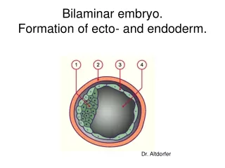
Inner cell mass develops into bilaminar embryonic disc • Hypoblast– Yolk sac forms here • Epiblast– amniotic cavity forms here • Yolk sac– formed by an extension of hypoblast • Digestive tube forms from yolk sac • Tissues around yolk sac • Gives rise to earliest blood cells and blood vessels • Amniotic sac – formed by an extension of epiblast • Outer membrane – forms the amnion • Inner membrane – forms the amniotic saccavity • Filled with amniotic fluid
2- (bilaminar germ disc) The inner cell mass is differentiated into: a- hypoblast = endoderm (small cuboidal cells facing the blastocyst cavity and b- epiblast= ectoderm(tall columnar cells facing the amniotic cavity).
Formation of Amniotic Cavity As implantation of the blastocyst progresses, changes appear in the inner cell mass (embryoblast) A cavity, amniotic cavity appears separating embryoblast from the trophoblast, which soon becomes lined by amnioblasts derived from inner cell mass The cavity gradually increases in size and is filled with amniotic fluid
Formation of Primitive Yolk Sac The blastocyst cavity becomes lined with exocelomic membrane and is called exocelomic cavity The hypoblastic cells soon replace the exocelomic membrane and the cavity is then named as the primitive (primary) yolk sac
3- FORMATION OF THE EXTRA-EMBRYONIC MESODERM AND EXTRAEMBRYONIC COELOM - Cells appears between the inner surface of the cytotrophoblasts and the outer surface of the yolk sac cavity a. These cells are derived from the yolk sac cells and form the extra-embryonic (primary) mesoderm. b. They fill the space between the trophoblasts, the amnion and yolk sac cavities.
Large spaces develop in the extraembryonic mesoderm and coalesce to form extraembryonic coelom. These spaces rapidly fuse to form a large fluid filled, C-shaped cavity, the extraembyoniccelome surrounding the amniotic cavity and the yolk sac
Formation of Connecting Stalk The region where no cavity has appeared, forms the connecting stalk, that connects the amniotic cavity, yolk sac and the embryonic disc to the outer wall The site of the connecting stalk determines the caudal pole of the embryonic disc
With the formation of extraembryonicceolum: The extraembryonicmesoderm splits into two layers: an outer extraembryonic parietal (somatic) mesoderm an inner extraembryonic visceral (splanchnic) mesoderm
Clinical application: • The syncytiotrophoblast secretes (human chorionic gonadotrophinhormone) HCG. • It stimulates the production of progesterone which in turn is important in sustaining the placenta. By the end of the 2ndweek • the amount of this hormone HCG will be sufficient to be detected in the maternal blood and urine. This is the basis of pregnancy test.
EXTRAEMBRYONIC MESODERM Cells are derived from epiblast and hypoblast 2 layers: Somatopleuric mesoderm- lining the amnion Splanchnopleuric mesoderm- lining the yolk sac Free template from www.brainybetty.com
The primary yolk sac decreases in size and becomes the secondary (definitive) yolk sac Wall of the yolk sac, amnion & chorion are formed
The second week of development is the week of two`s, because of the following: The trophoblast differentiates into 2 layers, cytotrophoblast& syncytiotrophoblast The inner cell mass differentiates into 2 layers, epiblast& hypoblast. The primary mesoderm splits into somatopleuric primary mesoderm & splanchnopleuric primary mesoderm. Starting of formation of the amniotic and yolk sac cavities.