Download
1 / 39
470 likes | 1.06k Vues
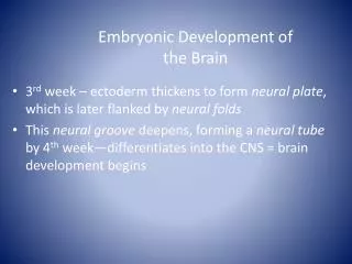
Formation of The Bilaminar Embryonic Disc THE SECOND WEEK. Prof.Dr.Meltem ÖZGÜNER Department of Histology-Embryology. THE SECOND WEEK. Trophoblast Embryoblast Extra - embryonic mesoderm At the end of the second week - Primitive uteroplacental circulation - Bilaminar embryonic disc

E N D
Formation of TheBilaminarEmbryonicDiscTHE SECOND WEEK Prof.Dr.Meltem ÖZGÜNER Department of Histology-Embryology
THE SECOND WEEK • Trophoblast • Embryoblast • Extra-embryonicmesoderm At theend of thesecondweek -Primitiveuteroplacentalcirculation -Bilaminarembryonicdisc -Theprochordalplatedevelops
IMPLANTATION • Implantation of theblastocystbegins at theend of thefirstweekand is completedbytheend of thesecondweek
IMPLANTATION Theprocessmay be summarized as follows: • Day 5:The zona pellucidadegenerates . Itsdisappearanceresultsfromtheenlargement of theblastocyst. • Day 6: Theblastocystattachestoendometrialepithelium.
IMPLANTATION • Day 7: Thetrophoblastbeginstodifferentiate in totwolayers, syncytiotrophoblastandcytotrophoblast. 1-thecytotrophoblast, a layer of mononucleatedcellswhich is mitoticallyactiveandformsnewcellsthatmigrateintothesyncytiotrophoblasts 2-thesyncytiotrophoblast, whichrapidlybecomes a large, thick, multinucleatedmass in which no cellboundariesarediscernible.
IMPLANTATION • Thesyncytiotrophoblastbeginstoproducehumanchorionicgonadotrophin (hCG), andenoughhCG is produced at theend of secondweektogive a positivepregnancy test. • hCGinducescorpusluteumforprogesteronsecretion.
IMPLANTATION • Day 8:Thesyncytiotrophoblasterodesendometrialtissues (capilleries, glands, stroma) andtheblastocyststartstoembed in theendometrium. • Day 9: Bloodfilledlacunaappear in thesyncytiotrophoblast.
IMPLANTATION • Day10:Theblastocystsinksbeneaththeendometrialepithelium. • Day 10 and 11: Lacunarnetworks form byfusion of adjacentlacunae.
IMPLANTATION • Day11 and 12: Thesyncytiotrophoblastcontinuestoerodeendometrialbloodvessels, allowingmaternalbloodtoseepintoandout of thelacunarnetworks, therebyestablishing a primitiveuteroplacentalcirculation
IMPLANTATION • Day 12 and 13: Thedefect in theendometrialepitheliumgraduallydisappears as thesurfaceepithelium is repaired. • Day 13 and 14: Primarychorionicvillidevelop
IMPLANTATIONDecidualReaction • Rapidproliferationanddifferentiation of thetrophoblastareimportantfeatures of thesecondweek of development. • Theseprocessesoccur as theblastocystimplants in theendometrium • Thevariousendometrialchangesresultingfromadaptation of thesetissuestoimplantation of theblastocystareknowncollectively as thedecidualreaction.
IMPLANTATIONDecidualReaction • Theblastocystattachtotheendometriallayer at theirembryonicpole; and at thisregion, trophoblastcellsdisplaceendometrialcells in thecentralpart of theimplantation site. • Thestromalcellsaroundtheimplantation site become laden withglycogenandlipidsandnamed as decidualcells. Theseprovide a richsource of embryonicnutrition.
AmnioticCavity, Amnion, BilaminarEmbryonicDiscandYolk Sac • Formation of theAmnioticCavityandAmnion • At thebeginning of thesecondweek, a smallcavityappears at theembryonicpolebetweentheembryoblastandtrophoblast.
Formationof theAmnioticCavityandAmnion • When it enlarges it becomestheamnioticcavity • Floor of theamnioticcavity is formedbytheepiblast
Formation of theAmnioticCavityandAmnion • Amnioblastsoramniogenic (amnion-forming) cells that separate from the epiblast and form a membraneknown as the amnion, which encloses the amniotic cavity.
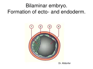
Formation of theBilaminarEmbryonicDisc • Concurrently, morphologicalchangesoccur in theembryoblastandresultswiththeformation of a flattened , bilaminarplate of cellscalledembryonicdisc.
Formation of theBilaminarEmbryonicDisc Thethickdiscconsists of twolayers: • 1-theepiblast, consisting of highcolumnarcellsrelatedtotheamnioticcavity, and • 2- thehypoblast, consisting of cuboidalcellsadjacenttotheblastocystcavity
Formation of theExocoelomicMembrane • At days 9 to 10, the cells from the hypoblast begin to migrate to the unembryonic pole forming a layer of cells just beneath the cytotrophoblast, called the Heuser's Membrane (or the exocoelomic membrane). • It surrounds the exocoelomiccavity.i.e. it lines the inner surface of the cytotrophoblast. • Heuser's membrane (or the exocoelomic membrane) is a short lived combination of hypoblast cells and extracellularmatrix.
Formation of theExocoelomicMembrane • At days 9 to 10, the cells from the hypoblast begin to migrate andform a thin layer of cells just beneath the cytotrophoblast, called the exocoelomic membrane (or the Heuser's Membrane).That is continuouswiththehypoblast.
Formation of theExocoelomicMembrane • It surrounds the blastocystcavitywhich is nowcalledtheexocoelomiccavityandit lines the inner surface of the cytotrophoblast. • Heuser's membrane (or the exocoelomic membrane) is a short lived combination of hypoblast cells and extracellularmatrix.
Formation of theExocoelomicMembrane • Theexocoelomicmembraneandcavitysoonbecomemodifiedto form theprimaryyolk sac (primitiveyolk sac) • Theembryonicdiscnowliesbetweentheamnioticcavityandtheprimaryyolk sac.
Formation of theExtraembryonicCavity • At days 11 to 12, cells, probablyfromthehypoblast, giveriseto a layer of loosely arranged cells that inserts aroundtheamnionandprimaryyolk sac, calledtheextraembryonicmesoderm.
Formation of theExtraembryonicCavity • As changesoccur in thetrophoblastandendometrium, theextraembryonicmesodermincreasesandisolatedcoelomicspacesappearwithin it. • Thesespacesrapidlyfuseto form a large, isolatedcavitycalledtheextraembryoniccoelom
Formation of theExtraembryonicCavity • Extraembryoniccoelom(fluid-filledcavity) surroundstheamnionandyolk sac exceptwheretheyareattachedtothechorionbytheconnectingstalk • Extraembryonic somatic mesoderm also forms the connecting stalk that is the primordium of the umbilical cord.
Formation of theSecondaryYolk Sac • At day 13, as theextraembryoniccoelomforms, theprimaryyolk sac decreases in size and a smallersecondaryyolk sacforms.It is oftenreferredtosimply as yolk sac.
Formation of theSecondaryYolk Sac • Secondaryyolk sac is formedbythecellsthatmigratefromthehypoblast inside theprimaryyolk sac. • Theyolk sac containsfluid but no yolk. • Itappearstohave a role in thenutrition of theembryonicdisc.
Blastocyst - day 13 1. Secondaryyolk sac2. Exocoelomiccyst3. Aminoticcavity4. Extra-embryoniccoelom5. Epiblast6. Connectingstalk7. Hypoblast8. Primaryvilli9. Trophoblasticlacunae10. Extraembryonicsomaticmesoderm11. Extraembryonicsplanchnicmesoderm Blastocyst - day 12 1. Syncytiotrophoblast2. Trophoblasticlacunae3. Surfaceepithelium4. Fibrin coagulum5. Epiblast6. Aminoticcavity7. Hypoblast8. Cytotrophoblast9. Exocoelomiccavity
DEVELOPMENT OF THE CHORIONIC SAC • Theend of thesecondweek (at approx. day 13) is characterizedbytheappearance of primitivechorionicvilli.
PRIMITIVE CHORIONIC VILLI • Proliferation of cytotrophoblastcellsproduceslocalmassesthatextendintothesyncytiotrophoblast.
PRIMITIVE CHORIONIC VILLI • Thecellularprojections form primarychorionicvilliwhichrepresentthefirststage in thedevelopment of thechorionicvilli of theplacenta.
DEVELOPMENT OF THE CHORIONIC SAC Theextraembryoniccoelomsplitstheextraembryonicmesodermintotwolayers: • 1-extraembryonicsomaticmesoderm, liningthetrophoblastandcoveringtheamnion • 2-extraembryonicsplanchnicmesoderm, surroundingtheyolk sac.
DEVELOPMENT OF THE CHORIONIC SAC Theextraembryoniccoelomsplitstheextraembryonicmesodermintotwolayers: • 1-extraembryonicsomaticmesoderm, liningthetrophoblastandcoveringtheamnion • 2-extraembryonicsplanchnicmesoderm, surroundingtheyolk sac.
CHORION Theextraembryonıcsomatıcmesoderm and twolayers of trophoblastconstitute CHORION
CHORIONIC CAVITY • Thechorionformsthewall of thechorionic sac, withinwhichtheembryoanditsamnioticandyolksacsaresuspendedbytheconnectingstalk • Theextra-embryoniccoelom is nowcalledtheCHORIONIC CAVITY
PROCHORDAL PLATE • The 14 dayembryostill has the form of a flat, bilaminarembryonicdisc, but thehypoblasticcells in a localizedareaarenowcolumnarand form a thickened, circularareacalledtheprochordalplate.
PROCHORDAL PLATE • Thisplateindicatesthefuture site of themouthand is an importantorganizer of theheadregion.