Embryonic Development of the Brain
630 likes | 834 Vues
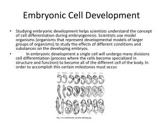
This overview details the embryonic development of the brain, beginning in the 3rd week with the ectoderm thickening to establish the neural plate, eventually leading to the formation of the neural tube by the 4th week. The subsequent development includes the emergence of the primary brain vesicles—the forebrain, midbrain, and hindbrain—and their subdivisions. Key structures such as the cerebral hemispheres, brain stem, basal nuclei, and various functional areas are explained, highlighting their roles in sensory and motor functions, memory, speech, and emotional processing.

Embryonic Development of the Brain
E N D
Presentation Transcript
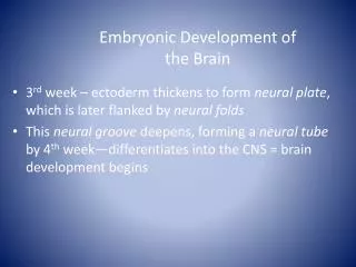
Embryonic Development of the Brain • 3rdweek – ectoderm thickens to form neural plate, which is later flanked by neural folds • This neural groove deepens, forming a neural tube by 4th week—differentiates into the CNS = brain development begins
Embryonic Development of the Brain • Between ectoderm and neural tube a neural crest forms, and the 3 primary brain vesicles appear: 1. FOREBRAIN, or prosencephalon[pros-en-sef-uh-lon] 2. MIDBRAIN, or mesencephalon[mes-en-sef-uh-lon] 3. HINDBRAIN, or rhombencephalon[rom-ben-sef-uh-lon] • The rest of the neural tube becomes the spinal cord
Embryonic Development of the Brain—figure 12.3 • Week 5 – Secondary brain vesicles arise • Forebrain divides: telencephalon[tel-en-sef-uh-lon, -luhn] (“endbrain”) and diencephalon [dahy-en-sef-uh-lon] (“interbrain”) • Hindbrain constricts: metencephalon[met-en-sef-uh-lon] (“afterbrain”) and myelencephalon[mahy-uh-len-sef-uh-lon](“spinal brain”) • Eventually the endbrain sprouts two lateral swellings – like Mickey’s ears • This eventually becomes the cerebrum • The other brain structures form the midbrain, pons, cerebellum and the medulla oblongata [ob-lawng-gah-tuh] • All but the cerebellum form the brain stem
Brain continues to grow rapidly; positions change • Midbrain & cervical flexures develop • Surfaces crease & fold = convolutions • Increase surface area = more neurons
Brain--major parts • Brain stem- continuous with spinal cord • Medulla oblongata, pons, midbrain • Diencephalon [dahy-en-sef-uh-lon] - above brain stem • Thalamus, hypothalamus & pineal gland • Cerebrum- at top and largest part • Surface covered with gray matter- cortex • Beneath is cerebral white matter • Cerebellum- back of brain stem • Means “little brain” • Cranial meninges[mi-nin-jeez]- dura mater, arachnoid[uh-rak-noid] mater & pia mater
Ventricles of the brain-filled with cerebrospinal fluid • Lateral ventricles, one deep within each hemisphere are large C-shaped chambers • They are separated by a thin membrane, the septum pellucidum • Interventricular foramen allows comminucation between the lateral ventricle and the narrow third ventricle • The third and fourth ventricles are connected by the canal-like cerebral aqueduct
The Cerebral Hemisphere • 3 basic regions: • Cerebral cortex • Internal white mater • Basal nuclei (gray matter within white matter) • Surface folds = gyri • Shallow grooves = sulci • Deeper groovesthat separate brain= fissures • Longitudinal Fissure- divides it into left & right hemispheres • Connected by corpus collosum • Transverse fissure- separates cerebral hemispheres from cerebellum
Cerebrum- Structure (cont) • Each hemisphere has 4 lobes • Frontal, parietal, temporal, occipital • Central sulcus separates frontal & parietal • Precentralgyrus anterior to sulcus= primary motor area • Postcentralgyrus = primary sensory area • Each hemisphere is concerned with the sensory and motor functions of the opposite side of the body. • The two hemispheres are not equal in function.
Function areas of Cortex • Specialized areas anatomically located • Sensory areas receive input and responsible for perception • Motor areas- initiate movements • Associative areas- complex integration: e.g. memory, emotion, reasoning, etc.
Motor Areas • Mainly from anterior part of hemisphere • Primary motor area- precentral gyrus • Broca’s speech area- • interacts with premotor area & primary motor area to regulate breathing and speech muscles
Sensory Areas • Primary somatosensory area-postcentralgyrus. • input includes: touch, proprioception, pain, itching, tickle, temperature • Primary visual area- occipital lobe • Primary auditory area- temporal lobe • Primary gustatory area – base of postcentralgyrus • Primary olfactory area- medial aspect of temporal lobe
Sensory Pathways • Relay information from periphery to cerebral cortex • 3 neurons in each pathway. • Posterior column- medial lemniscus[lem-nis-kuhs]pathway • Fine touch- body location, texture, size • Proprioception-[proh-pree-uh-sep-shuhn] position & motion of body parts • Vibratory sensations- fluctuating touch stimuli
Association Areas • Adjacent to sensory & motor areas • connected with tracts- interpret information • E.g. somatosensory[suh-mat-uh-sen-suh-ree] association area • Posterior to primary somatosensory area • Integrates sensation- exact shape & texture of object compares with stored memories • Wernike’s area- left temporal & parietal [puh-rahy-i-tl] lobes • Interprets meaning of speech • Right hemisphere adds emotional content
Lateralization • Left gets input from & sends output to right side of body and vice versa • Left important for spoken & written language, numerical & scientific skills & reasoning • Right more involved with spatial and pattern recognition and emotional content
Basal Nuclei • Deep gray = basal nuclei (basal ganglia) • Globuspalladus, putamen[pyoo-tey-min], caudate nucleus
Diencephalon • Thalamus- critical relay for sensory input • Transmits motor information from cerebellum & basal nuclei to cerebrum • Hypothalamus- important for homeostasis • Control of ANS-regulation of many activities • Control of pituitary and hormone production • Regulation of emotional & behavior patterns • Regulation of eating & drinking • Control of body temperature • Regulation of circadian rhythms & states of consciousness • Pineal gland- secretes melatonin
Brain Stem- Midbrain • Connects pons to Diencephalon • Large tracts = cerebral peduncles (motor) • Nuclei = substantianigra, rednuclei, cranial nerves III & IV • Superior colliculi – nuclei involved in tracking visual stimuli • Inferior colliculi – auditory input & startle reflex
Brain Stem- Pons • Pons (bridge)- nuclei & tracts • Connect left & right of cerebellum • Ascending & descending tracts • Nuclei – motor relays from cerebrum to cerebellum , respiration & cranial nerves V, VI, VII, VIII
Brain Stem- Medulla • Medulla Oblongata- inferior part of brainstem • white matter extending between spinal cord & other parts of brain • several nuclei: cardiovascular center • (heart rate) • Medullaryrhythmicity area • (respiratory rhythm) • Other sensory & reflex motor areas • Some related to cranial nerves
Cerebellum • Two cerebellar hemispheres • Posterior to medulla and pons, below cerebrum • Cerebellar cortex –gray matter • Tree like white matter & nuclei • Attached to brain stem via cerebellar peduncles
Cerebellar function • Gets wide range of sensory input • Compares with programmed motor activity from cerebral cortex • Smoothes & coordinates complex activities • Regulates posture & balance • Required for skilled motor activities
Limbic System • Ring of structures on inner border of cerebrum and floor of diencephalon • “emotional brain” – pain , pleasure, anger, affection, docility • Involuntary activity related to survival • Important in memory development
Reticular formation • Netlike arrangement of gray and white mater • Ascending part = Reticular Activating System (RAS) • Projects to cerebral cortex & helps maintain consciousness • Inactivation => sleep
Cerebrospinal Fluid (CSF) • Circulates through ventricles of brain and the subarachnoid [suhb-uh-rak-noid] space. • 4 ventricles: 2 lateral, third & fourth • Formed in choroid plexuses • = Specialized capillary networks in wall of ventricles covered by ependymal cells • Flows through ventricles then from 4th to central canal of spinal cord & subarachnoid cells • Reabsorbed through arachnoidvilli into superior saggital sinus
Brain blood supply • Requires ~20% body’s oxygen supply • 4 min lack => permanent damage • Requires continuous glucose supply • Protected by Blood-brain barrier • Allows lipid soluble materials: O2, CO2, alcohol, anesthetic agents but controls entry of other materials • Created by tight capillaries and glial cells
Sensory Pathways (cont) • Spinothalamic pathways- • anterior & lateral spinothalamic tracts • Relay impulses for pain, tickle, itch & thermal sensations.
Somatic Motor Pathways • Signals converge on lower motor neurons • Lower motor neurons stimulate muscles directly • Input comes from: • Local interneurons- e.g. reflexes • Upper motor neurons- corticospinal tracts • Basal ganglia- help with muscle tone • Cerebellum- coordination
Memory • Process for storing & retrieving information • Involves structural & functional changes • Involves association areas, parts of limbic system & diencephalon • Skill memory also involves cerebellum & basal ganglia
Cranial Nerves (table 9.6) • I Olfactory- sensory (smell) • II Optic- sensory (vision) • III Oculomotor- motor (eye) • IV Trochlear- motor (eye) • V Trigeminal- Mixed • Sensory around eyes & upper mouth motor to chewing • VI Abducens- motor (eye) • VII Facial- mixed • Sensory to front of tongue & motor to facial expression, lacrimal and some salivary glands
Cranial Nerves • VIII Vestibulocochlear- sensory (ear) • IX Glossopharyngeal- mixed • Sensory for rest of tongue, pharynx & palate, blood pressure • Motor to pharyngeal muscles, parotid salivary gland • X Vagus- mixed (major visceral nerve) • Sensory from pharynx, ear, diaphragm, visceral organs in ventral cavity • Motor to palatal & pharyngeal muscles & organs in ventral cavity • XI Accessory- Motor to voluntary muscles including sternocleidomastoid and trapezius • XII Hypoglossal- motor to tongue
Aging • Rapid growth during first few years • Size of neurons & proliferation of neuroglia increases • Increases development of dendritic branches & synaptic contacts • Decline in brain mass from early adulthood on
Spinal Cord StructureProtection and Coverings • Spinal cord in vertebral cavity- • Surrounded by bone • Wrapped in meninges- • 3 layers of connective tissue • Spinal cord meninges are continuous with brain meninges
Spinal Meninges • Epidural space lined with fat • Dura mater- tough ,dense connective tissue • Extends to 2nd sacral vertebra • Well beyond spinal cord • Arachnoid mater- collagen and elastic fibers • Subarachnoid space- • cerebral spinal fluid circulates in this space • Pia mater- transparent layer • adheres to surface of brain & spinal cord • Contains blood vessels
Gross Anatomy Of Spinal Cord • Runs to 2nd lumbar vertebra • Roots of spinal nerves for lumbar, sacral & coccygeal nerves in vertebral cavity before leaving = CaudaEquina • Enlargements: cervical & lumbar • Include nerves for upper & lower limbs • Each spinal segment gives rise to a spinal nerve – 31 pairs
Internal Structure Of Spinal Cord • Two grooves- left & right halves • Anterior median fissure & posterior median sulcus • Gray matter- 3 horns on each side • Anterior, posterior, lateral • Anterior- somatic motor neurons • Posterior- sensory neurons • Lateral- autonomic motor neurons