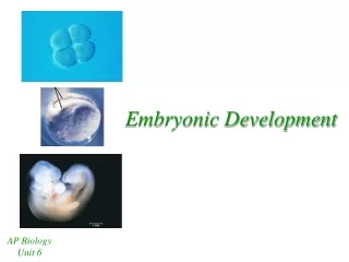
Embryonic Development
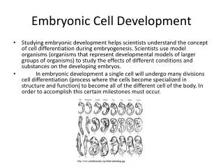
Embryonic Development. Variations in Embryonic Germ Layers and Body Cavity. Formation of the Digestive Cavity. At the Blastula stage, the embryo forms a fluid filled ball of cells


Embryonic Development
E N D
Presentation Transcript
Embryonic Development Variations in Embryonic Germ Layers and Body Cavity
Formation of the Digestive Cavity • At the Blastula stage, the embryo forms a fluid filled ball of cells • During Gastrulation, the blastula caves in on one end to form a Gastrula. The cavity formed by gastrulation will form a digestive cavity or digestive tract. • If gastrulation is incomplete, the gastric cavity will have only one opening. This arrangement is characteristic of Cnideria and Platyhelminthes • If gastrulation is complete, the digestive tract will have two openings. This arrangement has 2 variations, protostomes and deuterostomes
Protostomes vs. Deuterostomes In Protostomes, the blastopore forms the mouth. In Deuterostomes, the blastopore forms the anus and the mouth is the second opening
Embryonic Germ Layers • Organisms that undergo gastrulation fit into 2 categories: diploblasts and triploblasts • Diploblasts only have 2 embryonic germ layers • Gastrulation forms an inner layer of cells (endoderm) and an outer layer (ectoderm) • This diagram shows the endoderm in blue and the ectoderm in black
Embryonic Germ Layers • Triploblasts develop a third germ layer in between the endoderm and the ectoderm • The middle layer is called mesoderm • In the diagram, mesoderm is represented in yellow • Variations in the presence, absence and arrangement of mesodermal tissue is one of the most important distinguishing features of animal phyla
Phylogeny based on Mesodermal Arrangement • Phylum porifera lack a true digestive cavity • Cnideria are diploblastic. They have a digestive cavity but lack mesodermal tissue • The triploblastic phyla vary in regard to the arrangement of mesoderm and body cavity
Development of Mesodermal Tissue • Mesoderm is formed by a migration of endodermal cells into the blastocoel. • Mesoderm forms by one of 2 possible mechanism • The diagram at the right shows mesoderm development in acoelomates and protostomes • The formation of mesoderm initiates near the blastopore. Dividing mesodermal cells migrate into the blastocoel
Acoelomate phyla • Acoelomates do not form a body cavity • Flatworms (Platyhelminthes) are acoelomate • Mesodermal cells fill the entire blastocoel • Internal organs form, but are not separate and distinct
Pseudocoelomate Phyla • Pseudocoelomates have a body cavity, but mesoderm is associated with the outer body wall (ectoderm). • Nematodes and rotifers are pseudocoelomate • All pseudocoelomates have a true digestive tract (2 openings) and are protostomes (blastopore = mouth) • The digestive tract lacks mesodermal tissue, thus is not muscular
Cross Section of a Pseudocoelomate • The diagram shows a cross section through a nematode • Notice the space between organs (body cavity), but note that there is only mesoderm on the outer boundary, associated with the body wall. The Intestine is a simple layer of epithelium (no muscle) • The organs are separate and distinct, but the body cavity is “false” – not fully lined with mesoderm
Coelomate Protostomes • A true body cavity is fully lined with mesodermal tissue. • In coelomate protostomes, the body cavity is “schizocoelous” (note the descriptive root word “schizo”) • The solid mass of mesoderm filling the blastocoel splits due to the programmed cell death (“apoptosis”) of some centrally located cells • The diagram shows the coelom in pink
Coelomate Deuterostomes • All deuterostomes have a true body cavity • In deuterostomes, the blastopore forms the anus (so the “second opening” forms the mouth”) • In deuterostomes, the mesoderm forms from outpouching of the gut • Since it is “caving in” it is “enterocoelous” • Because the coelom is formed from pouches, it is completely surrounded by mesodermal tissue