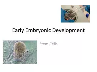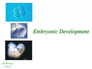
Embryonic development
Embryonic development. Ovum Fertilised ovum. Cell Division. Development of the embryo. Arm where an arm should be and not from the top of your head HOW? Fertilised egg fully formed neonate HOW?. Dolly the sheep. All nuclei are the same. All cells contain the same genes

Embryonic development
E N D
Presentation Transcript
Embryonic development • Ovum Fertilised ovum
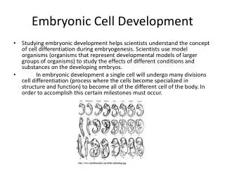
Development of the embryo • Arm where an arm should be and not from the top of your head • HOW? • Fertilised egg fully formed neonate • HOW?
Dolly the sheep All nuclei are the same • All cells contain the same genes • a complete copy of the genome • except gametes • Every cell with a nucleus can create every other cell in the body! – nuclear totipotency.
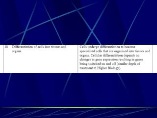
How's it controlled? Differential gene expression • Different cell types express (transcribe) only those genes needed to produce that tissue • i.e. only synthesises proteins needed e.g. muscle is only site of myoglobin production. • During development, genes are needed only at certain times, then switched off e.g. foetal haemoglobin • SPATIAL & TEMPORAL differential gene expression in development
Differential gene expression • Gene expression is regulated by • PROMOTERS & INHIBITORS (transcription factors) • Bind to regulatory sites near the genes and control transcription Animation
Differential gene expression • During development need to ensure correct promoters and inhibitors are present • Studied in drosophila
bicoid mRNA nanos mRNA The importance of the egg • Within the egg (before fertilization) a gradient of mRNAs is established • They code for proteins, that are transcription factors (known as morphogens) • Locate at either ends (the poles)
bicoid bicoid mRNA nanos mRNA nanos Distribution of proteins after fertilisation • Fertilisation stimulates the translation of bicoid and nanos mRNAs • The proteins diffuse • Set up a concentration gradient Egg Egg
First cell division • More bicoid than nanos protein • More nanos than bicoid protein
bicoid & nanos are transcription factors • bicoid and nanos regulate transcription of another set of genes • The segmentation genes (a class of genes which produce segments: GAP, PAIR RULE, SEGMENT POLARITY genes)) • They are also transcription factors • GAP controls PAIR RULE which control expression of SEGMENT POLARITY genes. • The SEGMENT POLARITY genes regulate expression of the homeotic genes – the final set of transcription factors. • Homeotic genes regulate expression of genes producing different parts of the body (i.e. structural proteins) This one gene controls many.
GAP GENE EXPRESSION Brief signals from a cascade of genes then split the fly embryo into ever smaller and many more specialized regions. In the photograph the embryo is divided into large blocks by proteins from so-called gap genes - Krüppel (red) and hunchback (green), which is turned on by bicoid 2½ hours after fertilization. The region where the two proteins overlap is yellow. The colors come from fluorescent dyes in antibodies that bind to these proteins.
PAIR RULE genes About a half hour later (3½ hrs), hairya "pair-rule" gene that is regulated by the gap genes, switches on and produces seven transient stripes. These stripes act like boundaries, dividing the embryo into seven segments
SEGMENT POLARITY genes • Finally, "segment-polarity" genes, divide each of the previous units into anterior and posterior compartments. • The narrow compartments correspond to specific segments of the embryo. • three head segments (H, top right), • three thoracic segments (T, lower right), • eight abdominal segments (a, from bottom right to upper right).
Segmentation genes divide the embryo into regions The drosphila embryo ends up with 17 segments Each segment will produce a different part of the body The instructions for the body parts are controlled by the HOMEOTIC GENES Animation
Homeotic gene expression determines ultimate function of segment • Bithorax mutant Mutant bithorax gene(s) Inappropriately expressed
Antennapodia complex mutant Mutant antennapedia complex gene(s) inappropriately expressed
In utero diethylstilbestrol (DES) exposure alters Hox gene expression in the developing mullerian system.Block K, Kardana A, Igarashi P, Taylor HS.Department of Obstetrics and Gynecology, Yale University School of Medicine, New Haven, Connecticut 06520, USA.Diethylstilbestrol (DES) was widely used to treat pregnant women through 1971. The reproductive tracts of their female offspring exposed to DES in utero are characterized by anatomic abnormalities. Here we show that DES administered to mice in utero produces changes in the expression pattern of several Hox genes that are involved in patterning of the reproductive tract. DES produces posterior shifts in Hox gene expression and homeotic anterior transformations of the reproductive tract. In human uterine or cervical cell cultures, DES induces HOXA9 or HOXA10 gene expression, respectively, to levels approximately twofold that induced by estradiol. The DES-induced expression is not inhibited by cyclohexamide. Estrogens are novel morphogens that directly regulate the expression pattern of posterior Hox genes in a manner analogous to retinoic acid regulation of anterior Hox genes. Alterations in HOX gene expression are a molecular mechanism by which DES affects reproductive tract development. Changes in Hox gene expression are a potential marker for the effects of in utero drug use that may become apparent only at late stages of development.
Summary • Maternal co-ordinate genes differentially distributed in the egg – they are transcription factors. • They regulate transcription of another set of genes • The segmentation genes (a class of genes which produce segments) • They are also transcription factors • After a cascade of 3 different types of segmentation genes (GAP, PAIR RULE, SEGMENT POLARITY), the homeotic genes are expressed • Homeotic genes are transcription factors – they regulate expression of genes producing different parts of the body • Each homeotic gene determines the anatomic fate of the area in which it is expressed.
VERTEBRATE DEVELOPMENT • In addition to differential gene expression, cell –cell communication and cell movements are important in the development of the vertebrate embryo. • Cells “talk” to neighbouring cells – organise the differentiation of their neighbours. • Cells migrate widely over the embryo.
CELL MIGRATION • Cells migrate towards diffusible chemical signals – chemotaxis • Along pathways of insoluble chemical - haptotaxis
Glycoproteins allow cells to adhere to each other and to the extracellular matrix