Download
1 / 28
350 likes | 892 Vues
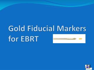
Gold Fiducial Markers for EBRT. Gold Fiducial Markers. Are used for soft tissue target volume localization and verification in external beam radiation treatment procedures like - IMRT 3D-confomal irradiation - CyberKnife or GammaKnive procedures - or even Brachytherapy

E N D
Gold Fiducial Markers Are used for soft tissue target volume localization and verification in external beam radiation treatment procedures like - IMRT • 3D-confomal irradiation - CyberKnife or GammaKnive procedures - or even Brachytherapy It might be used in organs like - Prostate - Liver - Pancreas - Brain
e.g. Prostate • EBRT nowadays is seen as one standard treatment for localized and advanced prostate cancer patients. • Prostate cancer is a multifocal tumor which causes the whole organ to be treated. The organ will be irradiated with homogeneous fields in 3D conformal technique or with IMRT. • Toxicity to bladder and rectum are limiting the dose to the organ to 72 Gray with regular irradiation procedures. • This table shows that higher doses are giving more local control. Source: Zelefski, Sloane Kettering CC, NY, NY
Intention Goal: High dose tothetargetvolume, lowtoxicitytotheorgansatrisk. But: The organsatriskareadjecenttothetargetvolume.
1. Approach to a solution The irradiatedvolumeshouldbesimilartotheshapeoftheorgantobetreated (conformal). • 3D – conformalirradiationshouldbethestandard. • Better but morecomplex: IMRT IntensityModulated Radiation Therapy • Fast moving leafs made from lead and controlled by a computer and shaping the beam from a LINAC. • The so called Multi Leaf Collimator. • Whit this system the contour of the beam can be shaped according to the shape of the organ at the respective angle of the radiation field. Conventional 3D conformal IMRT with organ tracking Source: Prof. D. Aebersold, Bern
But the main question is: Where is the Prostate ? Motion of the prostate during a 17 days cycle
Where is the Prostate ? • Organ motion and therefore geographic miss is a major problem when irradiating the prostate. • Hence a bigger target volume has to be irradiated. • But larger sections of the organs at risk will be irradiated as well. • This might lead to increasing toxicity rates.
Field edge variability during several treatment sessions Setup error of treatment sessions
Motion oftheprostate • Motion of the prostate might be induced by - Tension of the pelvis muscles, - Bowel and bladder filling - Breathing A cranial-caudal und ventral-dorsal misplacement of up to 2cm was observed. • Therefore conventional treatment scemesaks for a 1 – 2 cm margin around the organ. • Aside from that the prostate is shrinking during the treatment. Low margin low toxicity but at the risk not hitting the whole organ during each fraction local control ?? Big margin organ will be hit easily, but to save bladder and rectum the overall dose has to be reduced local control ??
Approach to a solution: IGRT • IGRT Image Guided Radiation TherapyRadiation Therapy with image guidance in intended to localize and adjust the target volume. The image is obtained by an on board imaging system (OBI). Today this can be a flat panel sensor with MV or kV imaging, extra portal imaging or cone beam CT. • BUT: • The prostate itself is hardly seen on x-ray images. • And bony structures might not be a reliable aid. Motion of the prostate versus bony structures
Approach to a Solution: Navigation aid = Marker • The marker should provide a reliable and reproducible localization of the prostate. • The marker should be easily seen with the OBI of the LINAC. • The marker has to be of a radio opaque material. E.g. gold, tungsten, silver etc., sometimes carbon. • It has to be biocompatible. Flourorefrence LINAC OBI
Why Gold • Gold does not reactwiththebody. • Gold is not causinganyallergies. • Gold provides a goodvisibilityunder x-rayandgamma-rays.
Whatis a goldmarkerandhowdoesitlooklike • Generally goldmarkersaresmallcylindersorballsmadeofgold. • Most commonlyusedsizesvaryfrom 0,8 to 1,2 mm in diameterand 3 to 5 mm in lenght . • Other dimensionsmightbeused in specialcases. • Goldmarker also canbemadeofthingoldspringsorsmallbeads on a suture.
Marker Kit The marker will be delivered sterile preloaded in a 20 cm needle blocked with bone wax or a synthetic spacer at the front end. It comes in sterile pouch ready for the implant.
Positioning of the Marker • To prevent the marker from migration or dislocation it has to be implanted in the organ. • Skin marker or patient immobilisation are unsufficient means.
Howmanymarkersareneeded • Usuallythreemarkers will beimplanted • In somespecialcasesfourmarkersmightbeneccessary. Thisadds extra confidenceandmighthelp in caseofdifficultbonyconditions. • Twomarkersareinsufficient.
Where to place the markers • Three markers will be place like • One at the Base one at the Apex and one most lateral in the middle of the gland. • OR: One on the left side at the base, one on the right side of the base and one under the urethra at the apex. • The markers have to be placed inside the gland 3 to 5 mm away from the capsule. • The actual position of the markers is not as important as the positioning at the maximum distance from each other.
Images Base – Middle – Apex
Images 4 Markers; Base – Base – Middle – Middle
Images Base – Base – Apex
How will the Markers implanted. • In many places the implant is performed by the Urologist under Ultrasound conrol. Usually he uses the biopsy channel of the rectal ultrasound probe to insert and visualize the needle. The implant is done through the rectal wall. • Some centers do have the equipment (stand, stepper, template grid) to perform a perineal insertion. This is more accurate but might cause a general anesthesia of the patient.
Procedure (one possible scenario) • In collaboration with the Urologists there will be a ultrasound guided implant of the Markers one or two weeks before the treatment planning starts. • With the localization of the Markers in the planning CT, the simulation and the daily treatment sessions the actual prostate position can be detected and the LINAC bed coordinates can be adjusted. This is the so called organ tracking. • Before each treatment session a control image is obtained and is matched with the reference image. • Prostate displacement will be compensated by moving the treatment bed according to the calculated geographic miss.
Procedure • ImplantofthemarkerbytheUrologist in theurologistsofficeorhospital. • Reference imagefortreatmentplanningandaquisitionofthemarkerposition • Postioningofthepatienton thebedofthe LINAC on thefirsttreatmentday.
Procedure • Aquisition of the images, • Each day in two orthogonal planes to detect the geographical miss. Modern LINACS offer a diagnsotic x-rayunit in a 90° angle tothe LINAC. Thisgivesthepossibiltytocreate x-rayimagesoreven CT scansbeforethetreatmentstarts. Digitallyreconstructed x-rayimageasreferenceforthemarkerposition (A) calculationofthecorrectionwith a goldmarkerimagefromthe LINAC.
Procedure • Calculation of the mismatch • Modern systems offer the possibility match the actual image with the reference image. The actual image will be aligned with the reference image on the computer monitor and the system generates the coordinates for the movement of the patients bed. • Correction of the bed • Modern systems do have a remote controlled bed. So compensation of the patient dislocation can be performed in an easy and fast way. Extra dose for imaging stays low. • Irradiation • Now the irradiation can start as usual. This localization and compensation procedure will be performed each day before each fraction. Mismatch Match
Conclusion • The prostate will be hit safely and reproducible during each treatment session. • Therefore a preferably high dose can be applied to the target volume. • Increase in local tumor control rate. • Decrease in toxicity.DieNebenwirkungsratewirdverringert. • Or: With similar toxicity the local control can be increased.
IGRT in pictures • Increased conformity and better dose distribution by means of IMRT. • Excellent match of the 95% iso dose level (blue area) with the target volume (red line). • The black arrows show the implanted gold markers. Gold marker Quelle: PIRUS GHADJAR, DANIEL M. AEBERSOLD