Download
1 / 14
140 likes | 268 Vues
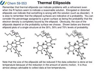
Thermal ellipsoids serve as indicators of potential issues in crystallographic refinement, even when R-factors suggest acceptable solutions. Elongated or distorted ellipsoids can point to structural problems, such as disorder. The size of these ellipsoids changes with the probability surface chosen, emphasizing the role of low-temperature data collection in reducing atomic motion. Additionally, understanding the SHELX list and CIF files is crucial for troubleshooting refinement processes and accurately reporting crystal structures, ensuring all pertinent information about atomic positions and displacement parameters is included.

E N D
Thermal Ellipsoids Remember that thermal ellipsoids can indicate problems with a refinement even when the R factors seem to indicate a reasonable solution. Elongated or distorted ellipsoids can indicate that something is wrong with the solution (such as disorder). It is wise to remember that the ellipsoid surfaces are indicative of a probability. You can consider the percentage assigned to a given surface as being the probability that the electron density is completely bound by the ellipsoid. Obviously, the size of the ellipsoids depend on the probability surface we choose. Shown below are thermal ellipsoid plots of a single structure at the 30%, 50% and 70% levels of probability. Note that the size of the ellipsoids will be reduced if the data collection is done at low temperature because of the reduction in the amount of atomic motion. It is thus almost always advisable to collect data at low temperatures.
Temperature Factors and Thermal Ellipsoids Remember that thermal ellipsoids can indicate problems with a refinement even when the R factors seem to indicate a reasonable solution. Consider the case of the molecule Cp*2SnMeX. With no H atoms attached to the Me groups, the following results were obtained when X was H, Li, C, or Cl. Notice that after a few cycles of refinement, the R1 factors for each of these solutions appears acceptable but the temperature factors and ellipsoids show that something is wrong for all of the options except X = C. R1 = 0.060: H3 2 0.591794 0.139842 0.156746 11.00000 0.00001 R1 = 0.048: LI3 5 0.592551 0.139622 0.156865 11.00000 0.01957
Temperature Factors and Thermal Ellipsoids R1 = 0.046: C3 1 0.592026 0.139458 0.156972 11.00000 0.04659 R1 = 0.056: CL3 4 0.589217 0.139459 0.157247 11.00000 0.26295 Consider why the size of the ellipsoid varies in this way: Since it is the electron density we are modeling, if there is not enough electron density provided by a given atom at a certain point, the only way to increase the electron density is to “shrink” the volume of space occupied by that atom. Conversely, if an atom provides too much electron density at a position in the cell, the only way to decrease the density is to increase the volume of space occupied by that atom.
The SHELX LIST file The a summary of the progress of each refinement cycle is stored in a file called name.lst (or shelxl.lst) and contains a variety of useful information. If your refinement has crashed for some reason, the list file can provide you with an indication of the cause of the problem. Likewise, if an error or warning message is provided at the end of the refinement, the name.lst file should offer information into the nature of the problem. For example, if we input a molecular formula that does not agree with the number and type of atoms found in our model, an error message will be produced. You will then find in the .lst file a statement such as: Unit-cell contents from UNIT instruction and atom list resp. C 140.00 140.00 H 156.00 0.00 B 4.00 4.00 N 4.00 4.00 F 60.00 60.00 Make sure that you understand why the numbers do not agree and then fix the problem. In this case, we haven’t included H atoms yet so none are found in the atom list. Other important information in the list file can be found in the correlation matrix (missed symmetry) and in the final listing of atomic positions (disorder).
The CIF file One of the most important files that we must generate after refinement is completed is the crystallographic information file (CIF) name.cif, which is the type of file that we submit when we report a crystal structure. Such files are created by inserting the ACTA command in the instruction files prior to the final refinement cycle. The name.cif file contains virtually all of the information that anyone might need to determine any desired information regarding the crystal structure to which the file refers. You should install a copy of the free enCIFer reader/viewer from the CCDC. I have given you a copy of a cif file for the structure of the ammonium borate as well as the tabulated data that are usually generated and reported. These tables generally include: 1. Crystal data and the refinement information. 2. Atomic coordinates, isotropic U values and their errors. 3. Bond lengths and angles and their errors (if desired torsion angles must be generated by the CONF command in the name.ins file) 4. Anisotropic displacement parameters Uij for the atoms. 5. Hydrogen atom positions, isotropic U values and their errors. You do NOT usually want to generate the list of calculated and observed structure factors – this was done in the past so that people could assess the data but now there are software (e.g. PLATON) and online methods (IUCr) of doing this.
Metrical Parameters The interatomic distances, angles between 3 atoms, torsion angles, planes and related quantities are known collectively as “Metrical Parameters”. Such parameters are readily calculated by software packages using the formulae provided in the “Derived Results” handout from Stout and Jensen. In many situations, we need to be able to compare these distances to other values that have been reported (e.g. to see if your bond lengths are typical or unusual). To do this, we must use the estimated standard deviations (ESD, si) associated with each particular parameter to assess if the quantities are the same (indistinguishable) or different to a certain level of probability. Comparing a group distances or angles: D = the difference in the distance or angle s = (Ssi2)1/2, l = D/s p is the probability that a quantity differs from the mean value by more than ls • p l • 1.00 0.000 • 0.50 0.674 • 0.10 1.65 • 0.05 1.96 • 0.01 2.58 • 0.001 3.2 These statistics are derived from normal distributions, where for example 99.7% of the time your result will be within 3s (3 standard deviations) of the mean value. Thus, a simple way to compare two values is to simply look at the range of values at ±3s. Thus a bond length listed as 1.522(3) Å indicates that there is a 99.7% probability that the bond is actually between 1.513 Å and 1.531 Å. If another bond length falls within this range, you can not distinguish between the two with a high level of confidence.
Metrical Parameters When you are reporting your crystallographic results, one of the most common points of discussion is the comparison of the metrical parameters in your structure to those of other structures. While the metrical parameters for other structures are generally given in the original literature, the best tools that we use to find the metrical parameters (and the original reports) are the various crystallographic databases. The databases are divided into compound types: Inorganic compounds containing no C-H bonds are found in the Inorganic Crystal Structure Database (ICSD). The department does not have a current license for this database but there is a free limited version available online at (http://icsd.ill.fr/icsd/). Small Organic, Organometallic and Inorganic compounds are collected in the Cambridge Structural Database (CSD; located at the Cambridge Crystallographic Data Centre http://www.ccdc.cam.ac.uk/). These are searched using the ConQuest software. Proteins and large biological molecules are collected in the Protein Data Bank (PDB). While most of the class does not generally search for this type of molecule, the file name.pdb is one of the most extensively used formats for the exchange of crystallographic data.
Inorganic Crystal Structure Database The ICSD contains the structural information for minerals. We are attempting to obtain a current version of this database but some information is available from the free version available online at (http://icsd.ill.fr/icsd/). When you select the link to the demo version, the screen that appears will look like this: Most of the search parameters are obvious – the links above each of the boxes will provide help regarding that search parameter – and the most useful ones for our use are searches based on the elements in the structure or the mineral name. For example, if we want to search for structures containing calcium an phosphate anions, we can enter “Ca P O” in the Elements box and hit search to produce:
Inorganic Crystal Structure Database The buttons above the list of results can then be used to obtain more information on any of the structures that have been listed.
Inorganic Crystal Structure Database For example, the “Reference” button will provide you with the article in which the data was reported and the “Details” button will give you most of the information you would likely want for the structure. You can also download various file types (such as CIF and RES) so that you can examine and manipulate the structure with one of the viewing programs that we have seen or will see (e.g. Diamond, ORTEP-3, GRETEP, PowderCell, etc.).
Inorganic Crystal Structure Database The “Pattern” button can be used to generate the powder X-ray diffraction pattern of the compound you have selected. We will talk about powder XRD later but this feature can obviously be used to confirm the composition of a solid you have studied using XRD.
Inorganic Crystal Structure Database The “Structure” button can be used to display the arrangement of the atoms in the structure either as a PDB/Chime (this is easier) type file or as a VRML file.
Cambridge Structural Database The CSD contains the structural information about most of the small molecule structures that most of you make or study. We have a site license for the software and you should be able to install a copy on one of the computers in your lab. The CSD contains more than 500,000 structures and is updated every few months. There are a number of TUTORIALS found under the “Help” menu and you should become familiar with most of the standard searching techniques. You can search the CSD by: Structure - drawing Peptide - name Author/Journal information Compound Name Elements Formula Space Group Unit Cell Z/Density Experimental All Text Reference Code (CSD number)
Cambridge Structural Database If you are searching metrical parameters, the best search method is by structure drawing because you can draw the type of structural details that are required. While the best way to learn how to use this software is practice (as always) I have a few hints for you and I’ll show you a few important things to remember. 1. Be aware of the bond-types you insert in your search structure – you may get false negative results if the CSD has it drawn differently! Draw your structure with as much flexibility as possible (e.g. “any” type of bond) when you are in doubt. 2. Use templates wisely (for the same reason as above). 3. Select multiple elements carefully to avoid too many hits. 4. Remember that you can put 2D and 3D restrictions on the model! -2D restrictions include the number of atoms bonded to a selected atom, the charge on a particular atom etc. -3D definitions let you search for distances, angles, planes, centroids etc. 5. Remember that you can look at the 3-D structures of the molecules in ConQuest or Mercury and you can export desired structures as CIF, SHELX, PDB and many other file types for viewing in other software.