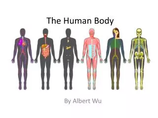
Understanding the Digestive System: Processes and Functions
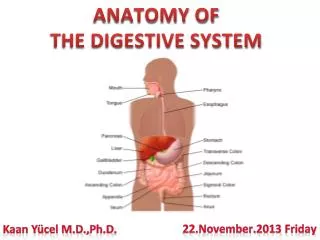
The digestive system is a complex network responsible for digestion, secretion, absorption, and motility within the gastrointestinal (GI) tract. It begins at the mouth and ends at the anus, with various organs and specialized cells that contribute to breaking down food and absorbing nutrients. Key functions include the secretion of digestive enzymes from the liver and pancreas into the duodenum, efficient water absorption, and coordinated motility for optimal nutrient absorption. Understanding these processes is essential for comprehending how our bodies extract and utilize essential nutrients.

Understanding the Digestive System: Processes and Functions
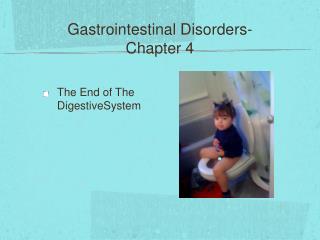
E N D
Presentation Transcript
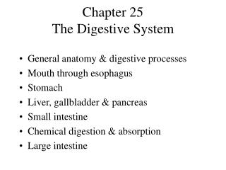
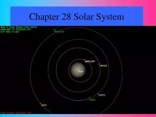
Chapter: 15 Digestive System
Figure 15-1 Digestion of food and absorption of nutrients are accomplished in a long tube connected to the external world at both ends; secretion and motility of “the tube” are major themes in understanding the gut.
Figure 15-2 The four processes carried out by the GI tract: digestion, secretion, absorption, and motility.
Figure 15-4 Digestive secretions from the liver and the pancreas are delivered into the duodenum of the small intestine through the sphincter of Oddi.
Figure 15-5 Digestive secretions are mostly water, with the average amounts indicated here. Note that only 100 ml are excreted in feces, so the mechanisms for water absorption are efficient (recall the kidneys’ primary role in water and osmotic homeostasis).
Figure 15-6 The gut wall has a layered organization, with the absorptive cells lining the lumen and neural and muscular components below. Blood and lymph vasculature is abundant to transport absorbed nutrients.
Figure 15-7 nutrients By projecting into the lumen, the villi increases the surface area for absorption of nutrients. Microvilli [aka brush border] fringe the villi to further increase surface area.
Figure 15-9 A molecular model of a bile salt, with the cholesterol-derived “core” in yellow. A space-filling model of a bile salt. The non-polar surface helps emulsify fats, and the polar surface promotes water solubility.
Figure 15-10 Bile salts and phospholipids convert large fat globules into smaller pieces with polar surfaces that inhibit reaggregation.
Figure 15-11 Emulsified fat globules are small enough that lipase enzymes gain access to degrade triglycerides to monoglycerides and fatty acids, which enter the absorptive cells by simple diffusion or aggregate to form loosely held micelles, which readily break down.
Figure 15-12 Big Droplets of Fat Small Droplets of Fat Micelles Fatty Acids and Monoglycerides Chylomicron Assembly Distribution and Processing
Figure 15-13 The enteric nervous system coordinates digestion, secretion, and motility to optimize nutrient absorption. Its activity is modified by information from the CNS and from local chemical and mechanical sensors.
Figure 15-14 The swallowing reflex is coordinated by the medulla oblongata, which stimulates the appropriate sequence of contraction and relaxation in the participating skeletal muscle, sphincters, and smooth muscle groups.
Figure 15-15 The coordinated sequence of contraction and relaxation in the upper esophageal sphincter, the esophagus, and the lower esophageal sphincter is necessary to deliver swallowed food to the stomach.
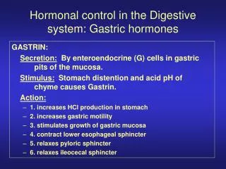
Figure 15-16 Specialized cells in the stomach synthesize and secrete mucous fluid, enzyme precursors, hydrochloric acid, and hormones. The abundant smooth muscle in the stomach is responsible for gastric motility.
Figure 15-17 Chief cells synthesize and secrete the protease precursor known as pepsinogen. Parietal cells synthesize and secrete the hydrochloric acid responsible for the acidic pH in the gastric lumen.
Figure 15-18 F O O D Acid production by the parietal cells in the stomach depends on the generation of carbonic acid; subsequent movement of hydrogen ions into the gastric lumen results from primary active transport.
Figure 15-19 One inhibitory and three stimulatory signals that alter acid secretion by parietal cells in the stomach.
Figure 15-20 Local and distant (CNS) information modulates the Enteric Nervous System’s activity regarding gastric motility and secretion.
Figure 15-21 The acidity in the gastric lumen converts the protease precursor pepsinogen to pepsin; subsequent conversions occur quickly as a result of pepsin’s protease activity.
Figure 15-22 Waves of smooth muscle contraction mix and propel the ingested contents of the gastric lumen, but only a small amount of the material enters the small intestine (duodenum) as a result of each wave cycle.
Figure 15-23 Rhythmic waves of smooth muscle contraction in the gut are the result of waves of action potentials moving along via gap junctions.
Figure 15-24 Delivery of acid and nutrients into the small intestine initiates signaling that slows gastric motility and secretion which allows adequate time for digestion and absorption in the duodenum.
Figure 15-25 The exocrine cells in the pancreas play a central role in the production of digestive enzymes; the endocrine functions of the pancreas will be discussed at length in Chapter 16.
Figure 15-26 Were digestive enzymes synthesized in their active form, they would digest the very cells that make them. Hence, inactive precursors (e.g., trypsinogen) become activated (trypsin).
Figure 15-27 Secretin’s receptors are found in the pancreas, which responds with additional bicarbonate delivery: gastric motility and secretion are inhibited.
Figure 15-28 • Cholecystokinin’s receptors are located: • in the pancreas, which • responds with additional • enzyme delivery • in the gallbladder, which • contracts to deliver more • bile • in the sphincter • of Oddi, which relaxes to • facilitate delivery of the enzymes and bile salts
Figure 15-29 Bile formation by cells in the liver includes 6 components: bile salts, lecithin, bicarbonate ions, cholesterol, bile pigments, and trace metals. The bile is funneled into the gallbladder and then delivered into the duodenum upon stimulation from CCK.
Figure 15-30 Up to 95% of the cholesterol-based bile salts are “recycled” by reabsorption along the intestine.
Figure 15-31 Cholecystokinin (CCK) stimulates the gallbladder, which responds by contracting and delivering more bile to the duodenum through the sphincter of Oddi, which relaxes (opens) in response to CCK.
Figure 15-32 Most of the contractions of the small intestine are of the mixing and churning actions portrayed here as segmentation contractions; peristalsis and the downstream movement of materials is infrequent.
Figure 15-33 In the large intestine, active transport of sodium, coupled with osmotic absorption of water, are the primary activities. Microbes here are active in the production of vitamin K.
Figure 15-34 Video endoscopy has greatly enhanced our understanding of normal processes in the gut, and reveals complications resulting from disease.