Download
1 / 72
730 likes | 1.36k Vues
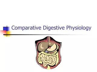
Digestive System General Functions of the Digestive System 1. Motility: a. Ingestion b. Peristalsis: rhythmic contractions moving food through the gastrointestinal tract. c. Defecation/excretion: removal the unabsorbed materials out of the body. 2. Secretion:

E N D
Digestive System General Functions of the Digestive System 1. Motility: a. Ingestion b. Peristalsis: rhythmic contractions moving food through the gastrointestinal tract. c. Defecation/excretion: removal the unabsorbed materials out of the body.
2. Secretion: Exocrine secretion: mucus, HCl, enzymes, H2O... - digestive juice, 2-3 L/day (70 % is reabsorbed). Endocrine secretion: hormones that regulate digestion activities.
3. Digestion: Mechanical and chemical decomposition of food. 4. Absorption: transport of digested products into blood and lymph. Lymphatic system: drain interstitial fluid and return it into veins, absorb lipid molecules from the digestive tract and send them to blood stream.
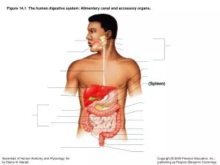
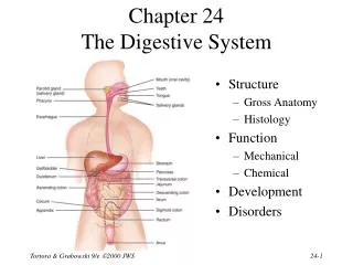
Anatomy: 1). Primary organs – gastrointestinal tract: Mouth pharynx esophagus stomach small intestines large intestines rectum anus (Food: Bolus chyme feces) 2). Accessory organs (2nd): Teeth, tongue, salivary glands, pancreas, liver and gall bladder.
Layers of GI tract: • Mucosa – lines the lumen of the tract and form • folds. It contains epithelial cells, connective • tissue, lymph nodules, capillary vessels and a • thin layer of smooth muscle. • Submucosa:capillary vessels, lymphatic tube, • glands and nerves (submucosal plexus).
Muscularis –inner circular and outer longitudinal layers of smooth muscles, responsible for segmental and peristaltic movement through the GI tract. Between the muscle layers: nerve fibers and ganglia of sympathetic and parasympathetic systems. Serosa– connective tissue, forms the very outside layer of the GI tract.
Functions of each organ: A. Mouth:(mechanical digestion) Saliva contains: H2O, mucus, and enzymes: Amylase: breaks down starch into disaccharides, Lysozyme: kills bacteria. Immunoglobulin - IgA: an antibody Functions of saliva: moistening food, dissolve chemicals…
Tongue: The mucous membrane of tongue contains taste buds, mucus glands, glands that secrete lipase. Inside of the tongue there are skeletal muscles that makes voluntary movement. Lipid digestion begins in the mouth by lingual Lipase. Functions of the tongue… participating swallowing.
B. Pharynx: Swallowing process is the result of corporation of tongue, larynx and pharynx. Tongue lifts up which causes the elevation of larynx to close trachea and open esophagus. So food enters the esophagus but not trachea. This is a precisely controlled process.
C. Esophagus The upper part contains skeletal muscle; the lower part contains smooth muscle. Skeletal muscle near pharynx perhaps participates in swallowing action. Lower esophageal sphinctercontracts after food passes into the stomach, which prevents the food from coming back to the esophagus.
D. Stomach Anatomy: Cardia - upper opening Stomach body Pylorus - lower opening Pyloric antrum - is the area near pylorus. Pyloric sphincter - Smooth muscles in the stomach body are oriented in different directions.
The epithelial cells of mucosa form gastric glands and secrete different products to form gastric juice. Goblet cells – secrete mucus; Parietal cells – produce HCL Chief cells – produce pepsinogen (a zymogen, inactive form of pepsin); ECL cells – secrete histamine and serotonin; G cells (endocrine cells) – gastrin;
Intrinsic factor required for the absorption of vitamin B12, is produced by parietal cells. Table 18.1
Gastric juice contains all the above substances and water. Food is mixed with gastric juice in stomach to form paste like substance called chyme.
Several important components in the gastric juice: Pepsin, a protease that cleaves peptide bonds and digests food proteins. It’s optimal working pH is 1-2. Pepsin is first synthesized as pepsinogen, which is cleaved and activated after it gets into the lumen of the stomach.
HCLis secreted by parietal cells. H+ is transported into the stomach against its concentration gradient by H+/K+ exchange pump. While H+ is pumped into the lumen of the stomach, K+ is taken up into the parietal cells.
Five functions of HCL: a. Kills microorganisms; b. Causes denature of proteins. c. Breaks down cell walls of plants; d. Activates pepsin, and provides an optimal environment for pepsin to function. Cooperative activities of pepsin and HCl allows the partial digestion of ingested proteins in the stomach. e. The acidic environment is also good for the absorption of Ca and Fe.
Mucus produced by Goblet cells, contains HCO3- and forms an alkaline coating to prevent the inner lining of stomach from being digested by HCl and pepsin.
Digestion and Absorption in the Stomach Proteins are partially digested in the stomach. The polypeptide chains are cleaved by pepsin into oligopeptides, tripeptides and dipeptides. There is very little digestion for carbohydrates and fats in the stomach. The major digestion process is in the small intestines.
Absorption in the stomach is very limited. The substances absorbed in stomach: alcohol, water, small amount of minerals (Ca, Fe, Na, Cl), and certain drugs, such as aspirin and anti-inflammatory drugs. The stomach empty time: for solid food is 4-6 hours; water - about 30 min. Pyloric sphincter controls the emptying speed of stomach. It also prevents the intestinal content from backing up into the stomach.
Regulation of The Stomach Function: (Nervous and hormonal controls) The movement and secretion of stomach are both stimulated by parasympathetic nerves. Cholinergic receptor blocker can decrease HCl secretion and smooth muscle contraction …
Locally produced hormone, gastrin, can also stimulate secretion and mobility of stomach. Secretion of gastrin is triggered by food entering the stomach. The effect of ACh and gastrin on gastric secretion is indirect. Both ACh and gastrin can stimulate ECL cells to produce histamine, which causes parietal cells to produce HCL.
Gastritis and peptic ulcer: Gastritis - inflammation in the stomach mucous membrane, can be induced by certain drugs (aspirin, steroids, anti-inflammatory drugs) and alcohol. Infection of a bacterium, helicobacter has been identified, as a direct cause of gastritis and gastric ulcer. Emotional and physical stress also contribute to gastritis.
Peptic ulcer or digestive ulcer, is open wound on the mucus membrane of the stomach or duodenum, often developed on the basis of gastritis. The mucous membrane of the stomach is digested by HCl and pepsin. General mechanism: combination of over production of gastric acid and underproduction of the alkaline mucus. (Helicobector is also involved.)
More specifically, duodenal ulcer may be more related with the over production of gastric acid. Gastric ulcer is often due to insufficient production of alkaline mucus. Gastric ulcer is closely related with stomach cancer.
The traditional treatment for digestive ulcer is acid control in combination with antacid drugs. Cholinergic nerve blocker to inhibit ACh release (Cimitidine). New generation: histamine receptors blocker: Zantag. Antibiotics are sometimes prescribed.
E. Small Intestines The major site for digestion and absorption The anatomy: Approximately 3 M long in a living person, but it will measure 6 M in a dead body, because of the relaxation of longitudinal muscles. Small intestines are held by mesentery - a broad sheet of connective tissue, which contains blood vessels lymphatic tubes and nerves.
Three segments: duodenum, jejunum, and ileum. • The walls of small intestines fold into a structure • called plica (plicae), which further folds into a • microscopic structure, villi. • The epithelial cells of each villi form smaller folds • microvilli. • Due to the folding plus villi and microvilli, the • total surface area is 2200 ft2, (otherwise 3.6 ft2)
Structure of Villus (Fig. 18.12) A finger-like fold of mucosa that projects into the intestinal lumen. Each villus is covered by columnar epithelial cells with goblet cells in between. In the center: connective tissue, capillary vessels, and a lacteal.
The absorbed carbohydrates and amino acids enter capillaries in the center of the villus, while lipids enter the central lacteal. Microvilli are brush like structure (brush border) formed by epithelial cells on the surface of villus.
Functions of small intestines: 1), Receiving the digestive juice from pancreas and liver (enzymes and bile salts). 2), Secretion: Intestinal juice, 1.8 L/day. It moistens chyme and neutralize it’s pH. Before absorption, pH of the food content must be neutralized.
a. NaHCO3 - is secreted by pancreas and small intestines. NaHCO3 Na+ + HCO3- H+ + HCO3 H2CO3 CO2 + H2O Duodenum is the major location of buffering action. Duodenum (10 inches long), receives chyme from stomach and neutralize it. The pH of chyme here changes from 1 to 7.5.
pH of the rest of the small intestines is ~ 7.5, which is optimal for most enzymes here. b. Enzymes Disaccharides, trisaccharides, dipeptides and tripeptides are further broken down by enzymes in the small intestines. Brush border enzymes stick to the cell surface and are exposed to the intestinal content.
Enterokinase is a brush border enzymes that activates trypsin that is produced by pancreas. Sucrase, maltase, and lactase are also produced by intestinal epithelial cells. (Lactase gene may stop expressing in adults.) Intestinal epithelial cells also make dipeptidase to cleave dipeptides into single amino acids which can then be absorbed.
c. Small intestines also produce hormonethat regulate secretion of stomach, pancreas and gallbladder. The arriving of chyme to duodenum stimulates the secretion of: Cholesytokinin (CCK) Chyme contianing lipids and proteins secretion of CCK, which stimulates contraction of gall bladder and secretion of pancreas.
3). Regulation: Arrival of chyme into duodenum also causes local production of secrenin, which stimulates the secretion of alkaline buffer, Na2HCO3, to neutralize the acidic chyme. Secrenin also inhibits gastric secretion and mobility.
4). Digestion and absorption The absorption of carbohydrate, lipids, Ca, Fe mainly occurs in duodenum and jejunum. Bile salts, water and electrolytes are absorbed in ileum.
Movement of small intestines: Segmentation is the major movement form in small intestines. Small, periodic, ringlike contractions cut chyme into segments and move it back and forth. Peristalsis is weak in the small intestines.
The smooth muscles in the small intestines are controlled by vagus. Smooth muscle cells have their own pace maker to produce slow rhythmic contraction even when there is no neural stimulation. Stimulation from parasympathetic system can accelerate both peristalsis and segmentation.
Diarrhea: If small intestines are overdistended or irritated, a strong peristaltic rush may pass through the entire length, sweeping the contents of the small intestines into the large intestines so quickly, that water, electrolytes and other substances that would normally be absorbed are excreted through frequent defecations.
F. Large intestines • Anatomy: • It has larger lumen than small intestines and • contains 4 parts: • 1.)Cecum, the beginning part of large intestines • and is a short blind pouch. • The nerrow tube hanging down - appedix. • Appendix is an immune organs, which contains • many lymphatic nodules and lymphocytes.
2). Colon: from right to left, it includes ascending, transverse, and descending colons. 3). Rectum 4). Anal canal. The main function of large intestines is the reabsorption of water and electrolytes. There is 1500 ml of materials enters the large intestines each day, but only 200 ml becomes feces.
Other functions of large intestines: Reabsorb and excrete bile salts. E.coli that normally residing in the large intestines produce vitamin K and some B vitamins, which can be absorbed in the large intestines.
G. Liver, Gall bladder andPancreas (accessory organs): Liver located at the upper right corner of the abdomen. Gall balder is attached to the liver between its two lobes.
Liver is full of blood supply. Liver cells are called hepatocytes, which form hepatic plates that are one to two cells thick. The spaces between hepatic plates are called Sinusoids, whichmimic capillary vessels. However, hepatic plates are more permeable. They have large pores and lack basement membrane, which allow passage of proteins, fat and cholesterol.
Within each hepatic plate, there are thin bile tubes that collects bile juice produced by hepatocytes. Kupffer cells ?
Hepatic portal system All blood leaving the small intestines is transported to liver through hepatic portal vein. The blood is filtered in the liver and then enters hepatic veins, which drains to inferior vena cava. Hepatic portal system: vein capillaries vein. The purpose is to extract nutrients and to remove toxins before they enter systemic circulation. (The liver also receives blood from hepatic artery.)
Functions of Liver • 1. Bile production and secretion • The liver produces 250 - 1500 ml of bile per day. • Major constituents of bile: bile pigments, bile • salts, phospholipids, cholesterol and ions. • Bile pigment includes billirubin and billiverdin. • Both are metabolic products of heme.