Cardiopulmonary Assessment & Pathophysiology Integration Program
This program demonstrates an orderly cardiopulmonary assessment integrated with cardiac anatomy and pathophysiology. Learn about the cardiac cycle events, auscultatory analysis, and cardiovascular evaluation methods to enhance bedside skills and patient care.

Cardiopulmonary Assessment & Pathophysiology Integration Program
E N D
Presentation Transcript
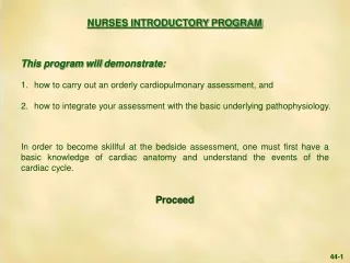
This program will demonstrate: 1. how to carry out an orderly cardiopulmonary assessment, and 2. how to integrate your assessment with the basic underlying pathophysiology. In order to become skillful at the bedside assessment, one must first have a basic knowledge of cardiac anatomy and understand the events of the cardiac cycle. Proceed NURSES INTRODUCTORY PROGRAM 44-1
BASIC CARDIAC ANATOMY Coronal section with pulmonary outflow tract, anterior ventricles and aorta cut away. Proceed AORTIC VALVE NON-CORONARY CUSP AORTA LEFT ATRIAL APPENDAGE RIGHT ATRIUM OSTIUM OF LEFT CORONARY ARTERY MEMBRANOUS INTRAVENTRICULAR SEPTUM MITRAL VALVE ANTERIOR LEAFLET CHORDAE TENDINEAE RIGHT ATRIAL APPENDAGE LEFT ANTEROLATERAL PAPILLARY MUSCLE TRICUSPID VALVE ANTERIOR CUSP TRICUSPID VALVE POSTERIOR CUSP LEFT POSTEROMEDIAL PAPILLARY MUSCLE CHORDAE TENDINEAE RIGHT POSTERIOR PAPILLARY MUSCLE LATERAL WALL LEFT VENTRICLE RIGHT ANTERIOR PAPILLARY MUSCLE WALL OF RIGHT VENTRICLE INTRAVENTRICULAR SEPTUM 44-2
120 AORTIC VALVE OPENS AORTIC PRESSURE 90 LEFT VENTRICULAR PRESSURE mm.Hg 60 LEFT ATRIAL PRESSURE MITRAL VALVE OPENS 30 0 SYSTOLE S2 S1 FIRST HEART SOUND SECOND HEART SOUND THE CARDIAC CYCLE Proceed Left ventricular systole begins with S1, that occurs when the pressure in the left ventricle rises above that of the left atrium (red arrow), closing the mitral valve. Systole ends and diastole begins with S2, that occurs when left ventricular pressure falls below that of the aortic root (blue arrow), closing the aortic valve. Analogous events of the cardiac cycle occur in the right heart, but at a lower pressure. 44-3
Blood flow is greatest early in systole and diastole. Approximately two-thirds of the blood ejected (stroke volume) leaves the ventricle in the first one-third of systole, and two-thirds of the blood filling the ventricle enters during the first one-third of diastole. This information is useful when analyzing the significance of certain auscultatory events. Proceed 44-4
When performing a cardiovascular evaluation, one should use an orderly method, such as the five finger approach advocated by Dr. W. Procter Harvey. THE FIVE FINGERS OF CLINICAL DIAGNOSIS We shall follow this outline, placing major emphasis on the physical assessment, especially auscultation. Proceed PHYSICAL ASSESSMENT ECG X RAY DIAGNOSTIC LABORATORY HISTORY 44-5
HISTORY 50-year-old man. CHIEF COMPLAINT:Although asymptomatic, his wife insisted he have a cardiac evaluation because her best friend’s husband recently had a heart attack. Question:What five key symptoms must be reviewed during a comprehensive cardiovascular history? 44-6
Answer: 1. Chest pain or discomfort e.g., ischemic cardiac pain or angina pectoris 2. Shortness of breath (dyspnea) at rest or with exertion e.g., as in left heart failure 3. Awareness of the heart beat (palpitations) e.g., tachyarrhythmia 4. Dizziness, lightheadedness or fainting e.g., syncope 5. Ankle swelling (edema) e.g., as in right heart failure Question: What additional general categories of the history should also be reviewed? 44-7
Answer: 1. Family history e.g., premature coronary artery disease, sudden death 2. Past history e.g., hypertension, diabetes, lipid abnormalities, murmurs 3. Social history e.g., smoking, alcohol, drug abuse The physical assessment can also be approached in a systematic manner as follows. 44-8
THE FIVE FINGERS OF PHYSICAL ASSESSMENT While auscultation is relegated to the last finger, it is the key assessment for nurses to carry out. The non-auscultatory findings will help to place the auscultatory assessment in context. We shall carry out the physical assessment in the order listed, beginning with the general appearance. Proceed PALPATE ARTERIAL PULSE INSPECT VENOUS PULSE PALPATE PRECORDIAL MOVEMENT AUSCULTATION INSPECT GENERAL APPEARANCE 44-9
PHYSICAL ASSESSMENT • GENERAL APPEARANCE – • The patient is normally developed and well nourished. He is comfortable, appears his stated age, and is neither acutely nor chronically ill. GENERAL APPEARANCE Question:How does the general appearance aid in evaluating a patient’s cardiac status? 44-10
Answer:The patient’s appearance can reflect both the state of the circulation and the presence of systemic disease that may also involve the heart. Examples of the state of circulation include: - cold, clammy skin and mental confusion, as seen in cardiogenic shock. - cyanosis and clubbing (shown below) as seen in a congenital right-to-left intracardiac shunt. Proceed 44-11
Answer (continued): Examples of systemic diseases include: - Marfan’s syndrome associated with aortic dissection. - Down’s syndrome (shown below) associated with an intracardiac shunt. Proceed 44-12
b. VENOUS PULSE - The next step in the physical assessment is the evaluation of the venous pulse. Information that may be obtained includes: - the central venous pressure (CVP) - the wave form VENOUS PULSE This is the least important cardiovascular assessment for nurses, especially the wave form. The venous pulse is evaluated by inspection of the transmitted internal jugular venous pulsations that directly reflect right atrial hemodynamics. They are observed (not palpated) as they undulate at the inferolateral aspect of the sternocleidomastoid muscle. Proceed 44-13
b. VENOUS PULSE (continued) A tangential light source can be used to better visualize the venous pulsations. Simultaneous palpation of the carotid artery will identify systole. Proceed 44-14
b. VENOUS PULSE (continued) - CENTRAL VENOUS PRESSURE The mean CVP is determined by measuring the vertical height of the venous pulsations above the mid right atrium, as the latter is the zero reference pressure. The patient should be examined in the semi-recumbent position at whatever angle the veins are best visualized. The sternal angle is used as a bedside reference point. It is 5 cm above the mid right atrium, and this relationship does not change significantly from the supine to the sitting position. Proceed 44-15
b. VENOUS PULSE (continued) - The CVP in our patient is estimated to be 3 cm H2O. The sternal angle is 5 cm above the mid right atrium. Since our patient’s neck veins undulate 2 cm below the sternal angle, his estimated CVP is 3 cm H2O. Proceed STERNAL ANGLE MID RIGHT ATRIUM 2 cm 5 cm 5 - 2 = 3cm 44-16
Neck veins normally do not pulsate to a level exceeding 2 cm above the sternal angle, i.e., the normal mean CVP is less than 7 cm. The most common cause of an elevated mean central venous pressure is an elevated right ventricular diastolic pressure, such as occurs with right ventricular heart failure. Proceed 44-17
VENOUS PULSE (continued) - WAVE FORM • Nurses will rarely need to address the venous wave form, but should know about the normal pattern shown. The first wave, due to right atrial contraction, occurs just before the carotid pulse (systole). The second wave, due to right atrial filling, occurs just after the carotid pulse. Atrial Contraction Atrial Filling S1 S2 SYSTOLE 44-18
ARTERIAL PULSE • ARTERIAL PULSE – • The next step in the physical assessment is the evaluation of the arterial pulses. This includes the measurement of heart rate and blood pressure. • In patients, the initial blood pressure should be taken in each arm using an appropriate size cuff that is tightly fitted. The cuff is inflated above systolic pressure and then slowly deflated. The level at which the first Korotkoff sound is heard is the systolic pressure. The level at which the Korotkoff sounds disappear is the diastolic pressure. Proceed 44-19
c. ARTERIAL PULSE (continued) - Our patient’s heart rate is 60 and his blood pressure is 120/80 mm Hg. Question:How do you interpret the patient’s blood pressure? Place the stethoscope diaphragm over the brachial artery and inflate the cuff. 44-20
Answer:The blood pressure is normal. In the adult, repeated resting blood pressures between 120/80 and 139/89 mm Hg are defined as “pre-hypertension.” As long as the patient has no symptoms, the lower the blood pressure the better. Proceed 44-21
c. ARTERIAL PULSE (continued) - CAROTID ARTERY Question:How do you interpret the carotid arterial pulse? S1 S2 44-22
Answer:The carotid arterial pulse is normal. The carotid artery is examined by placing the finger high in the neck between the trachea and the sternocleidomastoid muscle. Light pressure should be used to best evaluate the contour and to avoid cerebral ischemia. Proceed 44-23
For nurses learning the cardiopulmonary examination, the carotid pulse should be assessed. It will primarily be used for timing, as it identifies systole. The carotid artery is also assessed by evaluating: - rate of rise, e.g., normal, rapid or delayed - pulse volume, e.g., normal, large or small Proceed 44-24
A comparison of the normal pulse to an abnormal carotid arterial pulse is shown in the following graphic. The abnormal pulse is small in amplitude and slow in upstroke (hypokinetic). It is due to a decreased stroke volume, as may be seen with severe left ventricular failure. Proceed S1 S2 S1 S2 NORMAL ABNORMAL 44-25
PRECORDIAL MOVEMENT - • The next step in the bedside assessment is a systematic evaluation of precordial (chest wall) movements. First, inspect the general configuration of the thorax looking for obvious asymmetry or musculoskeletal abnormalities. Next observe the entire precordium for movement. PRECORDIAL MOVEMENT The palm of the hand is best suited for locating impulses and detecting vibrations. The fingertips are best for evaluating the size and contour of the impulse. Determine if the impulse is systolic or diastolic by simultaneously palpating the carotid artery or by listening to the heart sounds. Proceed 44-26
The precordial impulse should be assessed by analyzing: - Location - Size - Contour Palpation should be carried out in the areas shown on the diagram that follows. 44-27
d. PRECORDIAL MOVEMENT (continued) Proceed Most important for nurses are: a = APICAL = LEFT VENTRICULAR AREA (midclavicular line [MCL] 5th intercostal space [ICS]) b = DISPLACED APICAL AREA (anterior axillary line [AAL] 6th and 7th ICS) Less important is: c = RIGHT VENTRICULAR AREA (mid and lower left sternal edge) 44-28
d. PRECORDIAL MOVEMENT (continued) The only palpable impulse in this patient is at the apex. Question:How do you interpret the apical impulse? 5TH ICS MCL S1 S2 SYSTOLE 44-29
Answer: The apical impulse is normal. It is located in the fifth intercostal space, midclavicular line and is brief in duration, occurring immediately after S1. Such normal impulses are small (dime size). The apical impulse may be felt best with the patient in the left lateral decubitus position. Proceed 44-30
e. APICAL IMPULSE - In patients with left ventricular dilatation due to volume overload, e.g., aortic or mitral regurgitation, the apical impulse may be displaced inferolaterally and its size enlarged. Note the abnormal impulse is located in the anterior axillary line (inferolaterally displaced). It occupies the 6th and 7th ICS (enlarged) and is sustained throughout systole (abnormal contour). Proceed S1 S2 S1 S2 NORMAL 5th ICS MCL ABNORMAL 6th and 7th ICS AAL 44-31
Precordial movements may occur in diastole as well as systole. 5th ICS MCL In the abnormal tracing, there is a palpable presystolic impulse preceding the normal short systolic impulse. It reflects enhanced atrial contraction against a stiff or poorly compliant left ventricle. This impulse is common in patients with ischemic heart disease. Proceed S1 S2 S1 S2 NORMAL ABNORMAL 44-32
CARDIAC AUSCULTATION The final step in the physical assessment is auscultation. One must systematically listen in each of the classic acoustic areas shown below to evaluate the heart sounds and assess any murmur that might be present. Proceed AUSCULTATION - Aortic Area (Upper Right Sternal Edge) - Pulmonary Area (Upper Left Sternal Edge) - Tricuspid Area (Lower Left Sternal Edge) - Mitral Area (Apex) 44-33
We shall begin auscultation at the aortic area and move sequentially to the pulmonary, tricuspid and mitral areas. The diaphragm of the stethoscope is best for hearing high frequency sounds, and the bell is best for low frequency sounds. In each area, one should first listen selectively for the first and second heart sounds (S1 and S2), as these sounds respectively identify the beginning of systole and diastole. It is also helpful to palpate simultaneously the carotid artery to time the acoustic events. Both S1 and S2 are relatively high frequency and are, therefore, best heard with the diaphragm. Most important, the events of the cardiac cycle should be kept in mind when interpreting acoustic events at the bedside. Proceed 44-34
THE CARDIAC CYCLE In these left heart pressure tracings, S1 indicates the beginning of systole and coincides with closure of the mitral valve. S2 indicates the end of systole (and the beginning of diastole) and coincides with closure of the aortic valve. Proceed 120 AORTIC PRESSURE 90 mm.Hg LEFT VENTRICULAR PRESSURE 60 LEFT ATRIAL PRESSURE 30 0 SYSTOLE S1 S2 44-35
e. CARDIAC AUSCULTATION Question:How do you interpret the acoustic events at the upper right sternal edge? UPPER RIGHT STERNAL EDGE S1 S2 44-36
Answer: S1 is softer than S2. Both are single and normal in intensity. In this area, S1 is related to mitral valve closure and S2 to aortic valve closure. Both sounds are due to vibrations of the valve apparatus and surrounding cardiac structures and blood (“cardiohemic column”) that result from valve closure and the ensuing deceleration of blood flow. S2 is normally louder than S1 in the aortic area because the stethoscope is closer to the aortic valve than it is to the mitral valve. The relative intensities of the heart sounds may be clues to pathology. For example, a loud aortic closure (A2) may be seen in some patients with hypertension. 44-37
e. CARDIAC AUSCULTATION(continued) Question:How do you interpret the acoustic events at the upper left sternal edge? .04 A P UPPER LEFT STERNAL EDGE S1 S2 S1 Inspiration Expiration 44-38
Answer:There is normal inspiratory (physiologic) splitting of S2 due to asynchronous aortic and pulmonic closure of .04 seconds. A2 (aortic valve closure) is slightly more intense than P2 (pulmonic valve closure), as the aortic valve closes under a much higher pressure than the pulmonic valve. Inspiration causes a drop in intrathoracic pressure and an increase in lung volume. The result is greater venous return to the right ventricle and less return to the left ventricle. The increase in right ventricular volume prolongs right-sided ejection time, and the decrease in left ventricular volume reduces left-sided ejection time. In addition, inspiration allows the pulmonary arteries to receive a greater volume of blood, contributing further to a delay in P2. The net effect is a later P2 and an earlier A2. Proceed 44-39
Abnormal splitting of the second heart sound occurs in many pathologic conditions. An example follows: The second heart sound is widely split in both inspiration and expiration, i.e., “fixed splitting.” Fixed splitting is characteristic of a large atrial septal defect. Normal variation of S2 splitting does not occur in this situation because blood continuously flows left-to-right through the shunt, thereby counterbalancing respiratory effects. This results in constant, increased right ventricular flow. Proceed .07 .07 A P A P S1 S1 S1 Expiration Inspiration 44-40
e. CARDIAC AUSCULTATION(continued) Question: How do you interpret the acoustic events at the lower left sternal edge? .03 M T LOWER LEFT STERNAL EDGE S2 44-41
Answer:S1 is louder than S2, as the atrioventricular valves are closer to the stethoscope in this area than are the semilunar valves. There is normal splitting of S1 (.03 seconds) due to asynchronous closure of the mitral (M) and tricuspid (T) valves. In normal patients, the first heart sound may be single or split. Splitting of S1, if present, is best heard at the tricuspid area because the normally softer tricuspid component is best heard at this area. Proceed 44-42
e. CARDIAC AUSCULTATION(continued) Question:How do you interpret the acoustic events at the apex? APEX S2 S1 44-43
Answer:The heart sounds are normal. S1 and S2 are single and S1 is louder than S2. Other heart sounds may be heard at the apex. Most important are diastolic filling sounds, the third and fourth sounds (S3 and S4). Proceed APEX S4 S3 S2 S1 44-44
These low frequency diastolic filling sounds are best heard with the bell of the stethoscope. The S3 is related to the acceleration and deceleration of blood during early passive filling of the ventricle. It can be normal in children and young adults (enhanced acceleration during filling) but may also be present in heart failure (enhanced deceleration during filling). The S4 is related to atrial contraction filling a stiff or poorly compliant ventricle. Proceed 44-45
In some patients a murmur may be heard. A murmur is a sustained series of audible vibrations due to turbulent blood flow. Murmurs may occur in systole or diastole. They may also be continuous, i.e., continue through the second heart sound, as in patent ductus arteriosus. An example of a very common systolic murmur follows. 44-46
.05 A P UPPER LEFT STERNAL EDGE S1 Inspiration THE INNOCENT MURMUR This is a typical “innocent” murmur, often heard in youngsters. It is heard in early systole when the majority of blood leaves the ventricle, and is related to the normal turbulence of flow across the pulmonary outflow tract. Note there is also normal splitting of S2 during inspiration. Proceed 44-47
FOUR CLASSIC HEART MURMURS Murmurs may be characterized by some or all of the following descriptors: 1. Timing - phase of cardiac cycle - systolic, diastolic, continuous - location in systole/diastole - early, mid or late - duration in systole/diastole - short, long or throughout 2. Contour (shape) Crescendo Crescendo-Decrescendo Decrescendo Plateau 3. Frequency (pitch) - high - medium - low Proceed 44-48
MURMUR DESCRIPTORS (continued) 4. Quality blowing, harsh, rumbling, musical 5. Grade (loudness) 1 through 6 One is a murmur you can’t hear until you really concentrate, and six is amurmur you can hear with the stethoscope off the chest. 6. Location of maximal intensity 7. Radiation 8. Response of murmur to interventions respiration, changes in body position, Valsalva, etc. Proceed 44-49
.05 A P UPPER LEFT STERNAL EDGE S1 Inspiration Using these guidelines, the innocent murmur could be more fully described as short, early systolic, medium frequency, grade 2, crescendo-decrescendo and best heard at the upper left sternal edge. THE INNOCENT MURMUR Proceed 44-50