Download
1 / 15
150 likes | 183 Vues
Explore the basilar membrane vibration tuning curve and neural connections in the cochlea, encompassing afferent and efferent fibers of the auditory nerve. Delve into organization, discharge patterns, and encoding of frequency and intensity in auditory neurons.

E N D
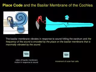
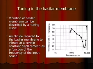
Tuning in the basilar membrane Vibration of basilar membrane can be described by a ‘tuning curve’ Amplitude required for the basilar membrane to vibrate at a certain constant displacement, as a function of the frequency of the input sound
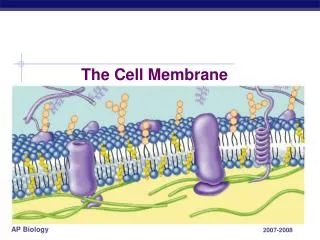
Neural connections in the cochlea Afferent and efferent fibers of the VIIIth cranial nerve (auditory nerve) Afferent: From organ of Corti to brain Efferent: From brain to organ of Corti Peripheral processes of auditory nerve neurons enter the cochlea through small openings on the edge of the osseous spiral lamina These openings are called ‘habenulae perforata’ These fibers are then gathered in the modiolus
Organization of the auditory nerve bundle in the modiolus Fibers from the apex in the middle Fibers from the base on the outside This nerve bundle then goes to the cochlear nucleus in the brain
Afferent fibers Around 30,000 neurons in man Only 5-15% of these innervate the OHC These are called Type II or outer spiral fibers One neuron connects to one OHC (one-to-many) Rest innervate the IHC These are called Type I or radial fibers Many neurons connect to one IHC (many-to-one)
Efferent fibers Originate from the olivocochlear bundle in the auditory brainstem Efferent fibers synapse on the afferent nerves innervating the IHC Efferent fibers synapse directly on the OHC
Discharge pattern of a neuron Neural spike has an initial large potential shift Following this, refractory or rest period of around 1 msec
Other terms Spontaneous discharge rate: Neuron’s discharge rate without a stimulus Threshold: Minimum stimulus level needed to cause an increase in the discharge rate above the spontaneous discharge rate
Spontaneous rate and thresholds Neurons with high spontaneous rate: Low threshold Neurons with low spontaneous rate: High threshold
Rate-Level function Also called input-output or intensity function Increase the level of the acoustic stimulus and measuring changes in the discharge rate of a single neuron
Response area Also called isolevel or isointensity curve Iso: “Same” Plotting how a neuron fires in response to sounds of different frequencies at a fixed intensity
Tuning curve Define a certain threshold for a neuron Plot the level of the tone required for this neuron to discharge at this threshold, as a function of the frequency of the tone
Encoding of frequency Two theories: Place theory Tonotopic organization Temporal theory Based on the periodic nature of nerve firing
Place theory Different neurons in the nerve respond to different frequencies Frequency of input determined by which neuron(s) in the auditory nerve fires at the greatest rate
Temporal theory For lower frequencies (< 5000 Hz), discharge rates of neurons are proportional to the period of the input stimulus. So for lower frequencies, rate of discharge of the neuron also provides information about the frequency of the stimulus.
Encoding of intensity Increase in discharge rate with increase in stimulus intensity However, for most neurons, increase in discharge rate only occurs for a limited range of input intensity Possible that discharge rate of many neurons may be combined to account for the 140 dB dynamic range of the ear