Download

1 / 24
250 likes | 865 Vues
Human Metaphase Chromosomes. Experiment Objectives. Preparing, staining and observing human metaphase chromosomes. Chromosome Morphology. Chromosomes are not visible under the light microscope in non-dividing cells (interphase cells).

E N D
Experiment Objectives • Preparing, staining and observing human metaphase chromosomes. Mazen Zaharna Molecular Biology 1/2009
Chromosome Morphology • Chromosomes are not visible under the light microscope in non-dividing cells (interphase cells). • As the cell begins to divide, the threads of chromatin (DNA-protein complex) in the nucleus begin to condense into multiple levels of coiled structures recognizable as chromosomes. • There are two modes of cell division: • mitosis and meiosis. Mitosis is responsible for the proliferation of body (somatic) cells, • whereas meiosis is responsible for the production of gametes. • Because mitotic cells are easy to obtain, morphological studies are generally based on mitotic metaphase chromosomes. Mazen Zaharna Molecular Biology 1/2009
Cell division • Cell division can be divided into: • Interphase, • Mitosis • Prophase, • Metaphase, • Anaphase, • Telophase. • Cytokinesis Mazen Zaharna Molecular Biology 1/2009
Metaphase • At metaphase the chromosomes are at their most condensed state, • Spindle fibers attaching to the area of the centromere called the kinetochore, forming pole-chromosome fibers. Mazen Zaharna Molecular Biology 1/2009
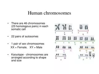
Chromosome Analysis • The best mitotic stage for chromosome analysis is prometaphase or metaphase. • A typical metaphase chromosome consists of two arms separated by a primary constriction or centromere. • Each of the two sister-chromatids contains a highly coiled double helix of DNA. Mazen Zaharna Molecular Biology 1/2009
Chromosome Analysis • Often the sister chromatids are so close to each other that the whole chromosome appears as a single rod-like structure • A chromosome may be characterized by its total length and the position of its centromere Mazen Zaharna Molecular Biology 1/2009
Types of Tissue • A variety of tissue types can be used to obtain chromosome preparations. • Some examples include peripheral blood, bone marrow, amniotic fluid and products of conception. • In the case of blood cell culture only cells that are actively dividing can be used for cytogenetic studies. • Normally only white blood cells are used for cytogenetic analysis. • Specific techniques differ according to the type of tissue used. Mazen Zaharna Molecular Biology 1/2009
Overview of Procedure • Collection of blood • Cell culture • Stopping the cell division at Metaphase • Hypotonic treatment of red & white blood cells • Fixation • Slide preparation • Staining Mazen Zaharna Molecular Biology 1/2009
1- Collection of blood • Draw 5 ml of venous blood into a sterile heparinized tube containing 0.1 ml of sodium heparin (500 units/ml). Mazen Zaharna Molecular Biology 1/2009
2- Cell Culture • Sterile technique must be used throughout the cell culture preparation, because it is possible to cause major contamination during this procedure • 70% of the problems are due to a lack of good sterile technique • Antibiotics do not eliminate problems of gross contamination which result from poor sterile technique or antibiotic-resistant mutants • Autoclaving renders pipettes, glassware, and solutions sterile Mazen Zaharna Molecular Biology 1/2009
2- Cell Culture Medium • Pipette 10 ml RPMI 1640 medium with L-Glutamine into a 15 ml labeled sterile culture tube • Supplement the medium with the following: Mazen Zaharna Molecular Biology 1/2009
2- Cell Culture Incubation • Add 1 ml of whole heparinized blood into the tube containing the supplemented medium • Mix contents of tube with gentle inversion • Incubate in 5% CO2 incubator at 37oC for 72 hours Mazen Zaharna Molecular Biology 1/2009
3- Stopping cell division at Metaphase • Pre-warm the Colchine (0.04 mg/ml) in incubator at 37oC • Add 25 µl of pre-warmed Colchine to the culture • Mix gently and incubate at 37oC for 30-60 minutes Mazen Zaharna Molecular Biology 1/2009
4- Hypotonic treatment of red & white blood cells • Centrifuge for 10 minutes at 2000 rpm • Discard supernatant without disturbing the cells leaving 0.5 ml of fluid • Add 1 ml of pre-warmed hypotonic solution (0.075 M KCl) at 37oC • Mix and then add 9 ml of hypotonic solution • Mix well by Pasteur pipette • Incubate at 37oC incubator for 17 minutes • hypotonic solution should not be in contact with cells more than 27 minutes (may cause rupture of WBCs) Mazen Zaharna Molecular Biology 1/2009
5- Fixation • Fixative must be prepared fresh • Add 3 parts of chilled absolute methanol: 1 part glacial acetic acid Mazen Zaharna Molecular Biology 1/2009
5- Fixation • Centrifuge for 10 minutes at 1000 – 1500 rpm • Remove supernatant leaving about 0.5 ml of fluid on top of cells • At this time there is probably a small whitish or reddish film at the bottom of the tube • The film contain red blood cell debris and enlarged WBCs Mazen Zaharna Molecular Biology 1/2009
5- Fixation • Add 5 ml of fixative to the tube • Mix with a Pasteur pipette 3-4 times • Place in refrigerator for 30 minutes • Centrifuge the tube for 10 minutes at 1000-1500 rpm • Remove supernatant and add another 6 ml of cold fixative, & mix well • Centrifuge the tube for 10 minutes at 1000-1500 rpm • Repeat the last two steps • Remove the supernatant leaving 1 ml of fluid at the bottom • The remaining material will be used to make the slides Mazen Zaharna Molecular Biology 1/2009
6- Slides Preparation • The slide must be exceptionally clean • Lay slides on a paper towel • Withdraw a few drops of cell suspension into a pipette • From a height of 20 cm, drop 2 or 3 drops of fluid on each slide • Allow the slides to dry Mazen Zaharna Molecular Biology 1/2009
7- Staining • Stain the slides by immersion in fresh Giemsa stain for 7-10 minutes • Remove slides from stain & rinse in distilled water • Observe under microscope X40 then under oil immersion Mazen Zaharna Molecular Biology 1/2009
http://www.biology.arizona.edu/human_bio/activities/karyotyping/patient_a/patient_a.htmlhttp://www.biology.arizona.edu/human_bio/activities/karyotyping/patient_a/patient_a.html • http://www.youtube.com/watch?v=E0WkZr819UU Mazen Zaharna Molecular Biology 1/2009