Cardiovascular Control During Exercise: Understanding Heart and Vascular System Function
650 likes | 779 Vues
This chapter explores the cardiovascular system's structure and function, focusing on how it meets increased demands during exercise. Learn how the heart, blood vessels, and blood work together to deliver oxygen and nutrients to active tissues, and remove waste products like carbon dioxide. The role of the autonomic nervous system in regulating heart rate, cardiac output, and the intrinsic conduction system is also discussed. Understand the significance of bradycardia and tachycardia, the cardiac cycle phases, and the mechanics of valve operation.

Cardiovascular Control During Exercise: Understanding Heart and Vascular System Function
E N D
Presentation Transcript
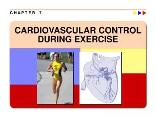
C H A P T E R 7 CARDIOVASCULAR CONTROL DURING EXERCISE
Learning Objectives w Review the structure and function of the heart, vascular system, and blood. w Find out how the cardiovascular system responds to increased demands during exercise. w Explore the role of the cardiovascular system in delivering oxygen and nutrients to active body tissues.
Cardiovascular System: What is it? w A pump (the heart) w A system of pipes (the blood vessels) w A fluid medium (blood)
Cardiovascular System: What does it do? w Delivery (e.g., oxygen and nutrients like glucose and FFA) w Removal (e.g., carbon dioxide and other by-products of metabolism) w Transportation (e.g., hormones, growth factors) w Maintenance of homeostasis (e.g., body temperature, pH) w Immunity (e.g., white blood cells, antibodies)
Cardiovascular System: Two Circuits • Systemic Circuit • Left side of heart • Pumps oxygenated blood to the whole body • Returns deoxygenated blood to right heart via veins • Pulmonary Circuit • Right side of heart • Pumps deoxygenated blood to the lungs • Returns oxygenated blood to left heart • Arteries: Away from heart • Veins: Towards heart • Oxygenated or Deoxygenated?
Myocardium aka Cardiac Muscle Tissue w Muscle thickness varies directly with stress placed on chamber walls. w Left ventricle has the larger muscle mass and can develop the higher pressures w With vigorous training, the left ventricle size increases. w Due to intercalated disks impulses travel from muscle fiber to muscle fiber in the heart (unlike skeletal muscle fibers)
Myocardium—The Cardiac Muscle w Intercalated disks allow the myocardium to act as “one large muscle fiber” so all fibers contract in a coordinated manner. Heart Muscle Skeletal Muscle Intercalated disk
Cardiac Conduction System • Sinoatrial (SA) node—pacemaker • Spontaneous contraction • Atrioventricular (AV) node • Slows conduction velocity and allows filling, so atrial contraction precedes ventricular contraction • AV bundle (bundle of His) • Transmits signal wPurkinje fibers - Rapid conduction that allows the ventricles to contract similarly.
Autonomic Innervation of the Heart Powers and Howley, Exercise Physiology, 2004
Extrinsic Control of the Heart • Parasympathetic nervous system acts through the vagus nerve to decrease heart rate. • The SA Node has an intrinsic firing rate of 100-110 bpm. • With innervation, RHR = 60-70 bpm • Ach is the primary nuerotransmitter.
Extrinsic Control of the Heart • Sympathetic nervous system is stimulated by stress (“fight or flight”) to increase heart rate. • Norepinephrine (NE) is the sympathetic neurotransmitter. • Upon stimulation, heart rate is increased. • Epinephrine and norepinephrine—released from adrenal medulla due to sympathetic nervous system activity.
Heart Transplant? • What happens when the nerves are cut? • Must rely on circulating catecholamine levels. • Resting Heart Rate is ?
Training Effects on Resting Heart Rate Resting heart rates in adults tend to be between 60 and 85 beats/min. However, extended endurance training can lower resting heart rate to 40 beats/min or less. This lower heart rate is thought to be due to increased parasympathetic stimulation. Referred to as the “athletic heart rate”
Cardiac Arrhythmias Bradycardia—resting heart rate below 60 beats/min; normal in trained persons (different pathology). Tachycardia—resting heart rate above 100 beats/min Premature ventricular contractions (PVCs)—Feel like skipped or extra beats; Ventricular tachycardia—Three or more consecutive PVCs that can lead to ventricular fibrillation (Vfib) in which contraction of the ventricular tissue is uncoordinated
Electrocardiogram (ECG or EKG) w Printout shows the heart's electrical activity and can be used to monitor cardiac changes and pathologies w The P wave—atrial depolarization w The QRS complex—ventricular depolarization and atrial repolarization w The T wave—ventricular repolarization
Cardiac Cycle w Events that occur between two consecutive heartbeats (systole to systole) wDiastole—relaxation phase during which the ventricles fill with blood (T wave to QRS)—62% of cycle duration wSystole—contraction phase during which the ventricles expel blood (QRS to T wave)—38% of cycle duration
Thought Question • What is the physical principle that dictates the opening and closing of the valves in the heart? • Hint: Think plumbing mechanics
. Cardiac Output (Q) • Total volume of blood pumped by each ventricle per minute. • Average cardiac output ~5 L/min • Cardiac Output = Stroke Volume x Heart Rate . • Q = HR ´ SV, e.g., Q = 60 beats/min x 75 ml/beat = 4,500 ml/min, or 4.5 l/min Cardiac Output
Stroke Volume • Stroke Volume (SV) • Volume of blood pumped by each ventricle per contraction. • End-diastolic volume (EDV) • Volume of blood in ventricle before contraction (Preload). • End-systolic volume (ESV) • Volume of blood in ventricle after contraction • SV = EDV – ESV, e.g., SV = 125 ml – 50 ml = 75 ml
Ejection Fraction (EF) w Proportion of blood pumped out of the left ventricle each beat • EF = SV/EDV x 100 • Averages 60% at rest, e.g., EF = 75 ml/125 ml x 100 = 60% • Often used as a measure of cardiac function
Vascular System: Closed System Plate from Andreas Vesalius’s De Humani Corporis Fabrica (1543) showing the vascular system.
Vascular System: Multiple Vessel Types • Arteries • Arterioles: Control blood flow through tissue by vasoconstriction and vasodilation • Capillaries: The site of exchange between blood and tissues occurs, e.g., oxygen, carbon dioxide, glucose, FFA, etc. • Venules • Veins: Contain valves to assist unidirectional flow of the blood
Vascular System: Closed System • Blood flows because of • A pressure difference, i.e., • a ΔP (93 mm Hg to 0) • Flow is unidirectional • because of valves in the • heart and the veins Ruch and Patton, Physiology and Biophysics, 1974
Blood Volume Distribution at Rest Note most of the blood volume is in the veins at rest, particularly in the viscera
Distribution of Cardiac Output (Q) w During steady state conditions, tissue blood flows are matched to the metabolic demands of the tissues wExtrinsic neural control — Sympathetic nerves innervating the smooth muscles within walls of vessels release NE, which generally causes the arterioles to constrict (alpha-adrenergic effect) wAutoregulation — With increased levels of metabolism, local vasodilator substances (e.g., K+) are released from the cells, overcoming the sympathetic vasoconstrictor effects, and causing the arterioles to dilate w Blood flow through a muscle is determined by the arterio-venous ΔP across the muscle and the radius of the arterioles
Blood Pressure w Systolic blood pressure (SBP) is the highest arterial pressure and diastolic blood pressure (DBP) is the lowest arterial pressure in the cardiac cycle • Mean arterial pressure (MAP)—average pressure exerted by the blood as it travels through arteries – usually what is considered in physiological studies • MAP = 1/3(SBP-DBP) + DBP • Usually around 93 mmHg
Blood Pressure ►Constriction increases blood pressure and dilation reduces blood pressure (garden hose effect) ► In the cardiovascular system: Pressure = Cardiac Output x Total Peripheral Resistance. • Total Peripheral Resistance = (Mean arterial pressure – mean venous pressure) / Cardiac Output • AKA the sum of all resistance in the systemic circulation. ►Mean arterial blood pressure is the primary regulated variable in the cardiovascular system, assuring sufficient blood flow to the brain in an upright bipedal human
Blood Pressure Regulation ► Monitored by baroreceptors located in the aorta and carotid arteries: - An increase in pressure → Results in Decreased sympathetic vasoconstriction and decreased inotropic effects on the heart. - A decrease in pressure → Results in Increased sympathetic vasoconstriction and increased inotropic effects on the heart
Blood w Blood is classified as a fluid connective tissue • Makes up about 8% of total body weight • So, a 70kg person would have ~5.6 liters of blood w Blood = Plasma + Formed Elements
Functions of the Blood w Transport gas, nutrients, wastes, and hormones w Regulates temperature w Buffers and balances acid-base
Blood Formed Elements and Hematocrit Blood: Formed Elements w White blood cells—Protect body from disease organisms w Blood platelets—Contribute to blood coagulation w Red blood cells – >99% of the total blood cells — contain hemoglobin, which binds and carries oxygen to tissues, and to a lesser extent, carbon dioxide to the lungs from the tissues Hematocrit • The percentage of blood volume that is made up of red blood cells.
Blood Viscosity w Thickness of the blood w The more viscous, the more resistant to flow • Higher hematocrits result in higher blood viscosity; the higher the hematocrit, the greater the oxygen carrying capacity; however, the greater viscosity requires greater cardiac work to pump the blood • What occurs during blood doping?
Cardiovascular System: During Exercise w What’s the goal? w Increase blood flow to active skeletal muscle in order to meet metabolic demands of the tissue. • Remember: Q = SV x HR • Therefore we need to increase Q
. • Stroke volume (SV) may increase up to 40% to 60% of maximal capacity before it reaches a plateau. • Increases in HR and SV during exercise allow cardiac output (Q) to increase. . Cardiovascular Response to Acute Exercise • Heart rate (HR) increases as exercise intensity increases up to maximal heart rate. w Blood flow increases to the active muscles, and decreases to the inactive tissue, e.g., visceral organs w Mean arterial pressure increases, but not to near the same extent as Q; thus, TPR decreases nearly proportionally to the increase in Q to maintain a constant MAP
Steady-State Exercise Heart Rate w Heart rate plateau reached during constant rate of submaximal work w Optimal heart rate for meeting circulatory demands at that rate of work w The lower the steady-state heart rate at a given exercise intensity (and the higher the stroke volume), the more efficient the heart (training effect).
Maximum Heart Rate w The highest heart rate value one can achieve in an all-out effort to the point of exhaustion w Remains relatively constant day-to-day but decreases with aging • Can be estimated: HRmax = 220 – age in years or
Stroke Volume w Determinant of cardiorespiratory endurance capacity at maximal rates of work because maximal HRs don’t vary much in persons of the same age w May increase with increasing rates of work up to intensities of 40% to 60% of max or higher when starting from upright position w May continue to increase up through maximal exercise intensity, generally in highly trained athletes
Stroke Volume Increases During Exercise wHOW IS STROKE VOLUME INCREASED? wFrank Starling mechanism — increase in venous return (i.e., increased pre-load) from muscle and respiratory pumps results in more blood in the ventricle causing it to stretch more and contract with more force according to the length-tension relationship. wIncreased ventricular contractility via increased sympathetic stimulation. wDecreased total peripheral resistance due to increased vasodilation of arterioles in active muscles (decreased afterload).
THE MUSCLE PUMP During exercise the muscle pump functions to return blood to the heart, or increase venous return; the thoracic pump serves the same function, i.e., to compress veins in the chest and abdomen to increase venous return to the heart
. CHANGES IN Q AND SV WITH INCREASING RATES OF WORK
w When exercise intensity exceeds 40% to 60%, further increases in Q may be more a result of increases in HR than SV since SV tends to plateau at higher work rates. . Cardiac Output w Resting value is approximately 5.0 L/min (70 kg male). w Increases linearly with increasing exercise intensity to maximal values of between 20 to 40 L/min. w The magnitude of cardiac output varies with body size.
. CHANGES IN HR, SV, AND Q WITH CHANGES IN POSITION AND EXERCISE INTENSITY