Download
1 / 41
430 likes | 619 Vues
Learn about dermatophytes Trichophyton, Epidermophyton, Microsporum, how they infect skin, and subcutaneous mycoses like Sporotrichosis and Chromoblastomycosis. Explore morphology, identification methods, epidemiology, and immunity factors.

E N D
SUPERFICIAL MYCOSES Assoc.Prof.Dr.Yesim Gürol
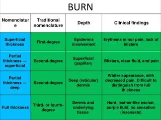
Learning Objectives • To list dermatophytes • To list subcutaneous mycoses
infections caused by • dermatophytic fungi (dermatophytosis) • Nondermatophytic fungi (dermatomycosis)
DERMATOPHYTOSES • Trichophyton • Epidermophyton • Microsporum • cause disease in animals and/or humans. • have the ability to invade the skin,hair or nails. • keratinophylic and keratinolytic. • invade upper outermost layer of epidermis
dermatophytosis tineas ringworm
Morphology and Identification identified by • colonial appearance • microscopic morphology • Growth at Sabouraud’s dextrose agar at 25oC for two weeks
Epidermophyton floccosum Microsporum canis Microsporum audouinii
Epidemiology and immunity • Begins in the skin after trauma and contact • Host susceptibility depends on • Moisture • Warmth • Spesific skin chemistry • Composition of sebum and perspiration • Youth • Heavy exposure • Genetic predisposition
Dermatophytes are classified as anthropophilic, zoophilic or geophilic according to their normal habitat. Anthropophilic • human hosts • mild, chronic inflammation. Zoophilic • primarily in animals • inflammatory reactions in humans who have contact with infected cats, dogs, cattle, horses, birds, or other animals. • followed by a rapid termination of the infection Geophilic • from the soil • occasionally infect humans and animals. • They cause a marked inflammatory reaction, which limits the spread of the infection and may lead to a spontaneous cure but may also leave scars.
Contagious • Frequently transmitted by exposure to shed skin scales, nails or hair containinh hyphae or conidia
Tinea capitis Tinea favosa Tinea corporis Tinea pedis Tinea manuum Tinea imbricata Tinea cruris Tinea barbae Tinea nigra Tinea ungium
Tinea capitis • infection of the scalp with a dermatophyte fungus. • Hair can be infected with Trichophyton (abbreviated as "T".) and Microsporum ("M".) fungi. classified according to how the fungus invades the hair shaft: Ectothrix infection • The fungal branches (hyphae) and spores (arthroconidia) cover the outside of the hair. • Ectothrix infections can be identified by Woods light (long wave ultraviolet light) examination of the affected area the vet uses this to check your cats fur. Endothrix infection • The hair shaft is filled with fungal branches (hyphae) and spores (arthroconidia). • Endothrix infections do not fluoresce with Woods light. Favus • caused by T. schoenleinii infection • honeycomb destruction of the hair shaft
When the hair is infected, • ectothrix • endothrix • favic
Tinea capitis may present in several ways. • Dry scaling – like dandruff but usually with moth-eaten hair loss • Black dots – the hairs are broken off at the scalp surface, which is scaly • Smooth areas of hair loss • Kerion – very inflamed mass, like an abscess • Favus – yellow crusts and matted hair • Carrier state no symptoms and only mild scaling
Trichophytid reaction (id reaction) • The patient may become hypersensitive to constituents or products of the fungus and develop allergic manifestations • Usually vesicles • Mostly on the hands • No fungi present in lesion
Tinea unguium (onychomycosis) Tinea ingualis (cruris) (jock itch)
SUBCUTANEOUS MYCOSES • Normally reside in soil or vegetation • Enter the skin or subcutaneous tissue by traumatic inoculation with contaminated material • Sporotrichosis • Chromoblastomycosis • Eumycotic mycetoma • Subcutaneous zygomycosis • Subcutaneous phaeohyphomycosis
SPOROTRICHOSIS • Sporothrix schenkii • Thermally dimorphic • Usually sporadic • Most common in warmer climates • Outbreaks related to forest work, mining, gardening • Classic infection traumatic inoculation of soil or vegetable or organic matter contaminated with fungus • Zoonotic transmission with armadillo hunters and infected cats
Chronic infection • Nodular and ulcerative lesions that develop along lymphatics • grossly may resemble a malignant process ‘squamous cell carcinoma’ • Dissemination to other sites rare !(bones,eyes,lungs, central nervous system)
Specimens: • Biopsy • Exudate from lesions • Culture
Chromoblastomycosis (chromomycosis) • chronic fungal infection • slow growing verrucous nodules or plaques • mostly in tropics • Pigmented fungi (dematiaceous fungi) • Fonsecaea • Cladosporium • Exophiala • Cladophialophora • Rhinocladiella • Phialophora
into the skin by trauma • Verrucous, wart like lesions • Cauliflower like nodules • Rarely elephanthiasis
Subcutaneous phaeohyphomycosis • Darkly pigmented septate hyphae in tissue • Cutaneous and systemic infections • Solitary encapsulated cysts in the subcutaneous tissue • Sinusitis • Brain abscesses (usually fatal) • All exogenous molds that normally exist in nature
phaeohyphomycosis • Exophiala, • Phialophora, • Wangiella, • Bipolaris, • Exserohilum, • Cladophialophora , • Phaeoannellomyces, • Aureobasidium, • Cladosporium, • Curvularia • Alternaria
MYCETOMA • Local swelling of infected tissue and interconnecting • Often draining • Sinuses or fistulae that contain granules • Actinomycetoma....mycetoma caused by an actinomycete • Eumycetoma....(Madura foot, maduromycosis) mycetoma caused by a fungus • Clinical features similar • Treatment different • Actinomycetomas more invasive
After traumatic inoculation with soil contaminated with one of these agents • Feet, lower extremities, hands and exposed areas • Suppuration, abscesses, granules, • Contagious muscle and bone • Deformation, loss of function • Very rarely foreign body (e.g.cardiac pacemaker) infection