Download
1 / 34
340 likes | 374 Vues
Learn about the master controlling system of the body, functions of the nervous system, its structural and functional classifications, neurotransmitters, nervous tissue anatomy, differences between CNS and PNS, and more.

E N D
Anatomy Ch. 7 Part 1 Introduction to The nervous system
Introduction • The nervous system is the master controlling and communicating system of the body. • Communication with body cells is done by using electrical impulses. • In order to maintain homeostasis the nervous system works with the endocrine system • The nervous system conducts impulses • The endocrine system uses hormones
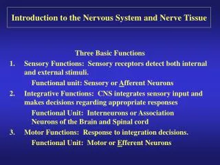
3 Functions of the Nervous System • Monitor changes produced by stimuli, also known as sensory input • Process and interpret, also known as integration • Cause a response by activating muscles or glands (effectors) via motor output
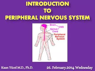
Structural Classification of the Nervous System • Central Nervous System (CNS) • Brain and spinal cord • Integrating and command center • Interprets and issues instructions • Peripheral Nervous System (PNS) • Spinal and cranial nerves • Link receptors to the CNS and the CNS to glands or muscles
Functional Classification (PNS) • Sensory (afferent) • Carry impulses to the CNS from sensory receptors • Divided into the Somatic and Visceral • Somatic: skin, skeletal muscles, and joints • Visceral: visceral organs • Motor (efferent) • Carry impulses from the CNS to muscles or glands • Divided into Somatic and Autonomic • Somatic: Voluntary control of skeletal muscles • Autonomic: involuntary control of smooth and cardiac muscles and glands
Sympathetic and Parasympathetic Divisions of the Autonomic Nervous System • Parasympathetic • Neurotransmitter is acetylcholine • Rest and Digest division • Decreases the activity of the cardiovascular and respiratory systems and increases the activity of the digestive system. • Sympathetic • Neurotransmitters are acetylcholine, epinephrine, and norepinephrine • Fight or flight division • Increases the activity of the cardiovascular and respiratory systems and decreases the activity of the digestive system.
Common Neurotransmitters • Acetylcholine • skeletal muscle activity • Adrenaline (epinephrine) • fight or flight • Noradrenaline (norepinephrine) • stress hormone • Serotonin • mood • Dopamine • pleasure • Endorphins • Euphoria (excitement or happiness) • Glutamate • memory • GABA • calming
Nervous Tissue • Supporting Cells (neuroglial cells) support insulate, and protect neurons • Astrocytes: link capillaries and neurons • Microglia: phagocytes that dispose of debris • Ependymal: circulate CSF • Oligodendrocytes: insulate nerve fibers • Schwann cells: form myelin sheaths • Satellite cells: protect and cushion
Anatomy of a Neuron • Cell body: nucleus, Nissl bodies (ER), and neurofibrils which maintain shape • Dendrites: convey incoming messages to the cell body • Axons: conduct messages away from the cell body
Regions of axons • Hillock: area of the cell body where the axon originates • Terminals: branches of axons that contain neurotransmitters • Synaptic cleft: gap between an axon and the next neuron, the junction is called a synapse
Myelin sheaths: covering of axons that protect and insulate and increase transmission rates of impulses • Schwann cells: produce myelin sheaths • Nodes of Ranvier: gaps between Schwann cells • Neurilemma: surrounds the myelin sheaths • Only found in the PNS which allows for regeneration
Differences between the CNS and PNS • In the CNS clusters of cells bodies are called nuclei • In the PNS clusters of cell bodies are called ganglia • In the CNS bundles of nerve fibers are called tracts • In the PNS bundles of nerve fibers are called nerves
The terms white matter and gray matter refer to regions of the CNS. • White matter has myelinated fibers • Gray matter has unmyelinated fibers
Functional Classification • Direction of Impulses • Sensory (afferent): carry impulses from receptors to the CNS; the cell bodies are outside of the CNS • Free nerve endings – pain and temperature • Meissners corpuscles – touch • Lamellar corpuscles – deep pressure • Proprioceptors – muscles and tendons
Motor (efferent): carry impulses from the CNS to organs, muscles or glands; the cell bodies are found in the CNS • Interneurons (association): connect motor and sensory neurons; cell bodies are found in the CNS
Neurons have 2 major functional properties: • Irritability: respond to stimulus • Conductivity: transmit impulses
Structural Classification • Number of Processes • Multipolar • several extensions from the cell body • all motor and association neurons • most common • Bipolar • 2 extensions from the cell body • only found in the eye and nose
Unipolar • A single short process that divides into proximal and distal processes • Sensory neurons of the PNS • Axons conduct impulses toward and away from the cell body.
Conduction of a Nerve Impulse • A resting neuron is polarized meaning there are more positive ions on the outside than on the inside • The major positive ion on the inside of the cell membrane is Potassium • The major positive ion on the outside of the cell membrane is Sodium
Steps of Impulse Conduction 1. As long the neuron is in the resting state it will be inactive. This is called the polarized state. Most neurons are excited by neurotransmitters. 2. When the neuron is stimulated, sodium quickly diffuses into the neuron. This changes the polarity of the membrane and is called depolarization.
3. If the stimulus is strong enough, the depolarization will travel over the entire length of the neuron membrane. This is called action potential or nerve impulse. 4. As the impulse moves, the permeability changes. Potassium is now able to leave the neuron rapidly by diffusion. This causes the cell to become repolarized. There cannot be another impulse until the neuron is repolarized.
5. After repolarization the sodium and potassium are returned to their original locations by the Na/K pump. This is an active process requiring ATP.
The previous process occurs in unmyelinated fibers. Fibers that have myelin conduct impulses much faster. • The impulse actually jumps from one node to another skipping myelinated regions. This is called saltatory conduction.
Impulses do not travel from one neuron to the next. • Instead neurotransmitters cross the synapse between neurons and carry the signal.
Steps of transmission from one neuron to the next (electrochemical) 1. When the action potential reaches the axon terminal, calcium enters the terminal 2. The calcium causes vesicles in the terminal to release the neurotransmitter 3. The neurotransmitter moves across the synapse and binds to receptors on the next neuron 4. If there are enough neurotransmitters to stimulate the next neuron then an impulse can be conducted. 5. Once the next neuron has been stimulated the neurotransmitter is removed.
Reflexes • Most of what the body does everyday is programmed as reflexes. • Reflexes are rapid, predictable, and involuntary responses to a stimuli • The pathway of a reflex is called a reflex arc • Reflexes can be classed as somatic or autonomic. • Somatic: skeletal muscles • Autonomic: smooth and cardiac muscles and glands
All reflexes have 5 elements: • Sensory receptor: reacts to stimuli • Sensory neurons: send message to the CNS • Integration center: the CNS • Motor neurons: send message to the effector organ • Effector organ: the muscle or gland stimulated
Reflex arcs can have 2 or more neurons. • Additional neurons are called interneurons. • The more neurons, the longer the reflex takes to happen. • Spinal reflexes do not require the brain’s involvement. • Reflexes that involve the brain are those that require many types of information.