Download

1 / 70
710 likes | 780 Vues
Learn about the classification, epidemiology, pathogenesis, clinical features, diagnosis, and therapy of Pemphigus Vulgaris (PV), Pemphigus Vegetans, and Pemphigus Foliaceus. Understand the genetic predisposition, antibody targets, and differential diagnosis.

E N D
Autoimmune BullousDiseases Dr. Abdullah ALAKEEL Assistant Professor & Consultant Department of Dermatology KKUH
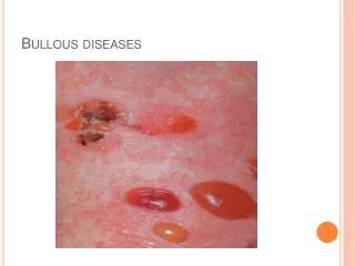
Circumscribed skin lesions containing fluid (If the size ≤ 5mm = vesicle If the size > 5mm = bulla)
Autoimmune bullousdiseases • A- Loss of intraepidermal adhesion: Pemphigus group : • 1- Pemphigus vulgaris (PV) with subtypes • a- Classic • b- Pemphigus vegetans 2- Pemphigus foliaceus with subtypes: a- Classic - Fogoselvagum - Pemphigus erythematosus ( Senear-Usher) b- Paraneoplastic pemphigus c- Drug induced pemphigus d- IgA pemphigus
Autoimmune bullousdiseases B- Loss of subepidermal adhesion : 1- Pemphigoid a- Bullous pemphigoid b- Pemphigoidgestationis c- Cicatricialpemphigoid 2- Linear IgA disease - of childhood - Adult form 3- Epidermolysisbullosaacquisita 4- Dermatitis herpetiformis
Autoimmune bullousdiseases • Pemphigus Group : • A group of disorders with loss of intraepidermal adhesion due to autoantibodies directed against proteins of the desmosomal complex that hold keratinocytes together. The desmosome is a complex structure, with many of its components targets for autoantibodies.
PV • Pemphigus vulgaris (PV): • Definition : severe, potentially fatal disease with intraepidermal blister formation on skin and mucosa caused by autoantibodies against desmogleins. • Epidemiology : 0.1-0.5/ 100000 yearly, most patients middle aged.
PV • Pathogenesis: • - Genetic predisposition: HLA-DRQ402- DQ0505 • - Antibodies against desmoglein 3 (Dsg 3) and later desmoglein 1 (Dsg 1 ). The bound antibodies activate proteases that damage the desmosome, leading to acantholysis. • - Serum antibody titer usually correlates with severity of disease and course.
PV - Agents containing sulfhydryl groups (penicillamine, captopril, piroxicam) are more likely to cause PV. - Those without sulfhydryl groups tend to cause PV ( beta-blockers, cephalosporins, penicillins, & rifampicin) . • Note : drugs from either group can cause either type of pemphigus.
PV • Clinical features: • Sites: oral mucosa, scalp , face, mechanicallystressedareas,nailfold, intertriginous areas. • Bliters are NOT stable, epidermisfallsapart, erosions & crusts are common • Oral involvement: 70%, anti-Dsg3 (Dsg 3 is the main desmoglein on mucosa) • Additionallocalizeddisease; scalp • Note:always check scalp whenconfrontedwithunexplained oral erosions.
PV • Generalizeddisease due to development of antibodiesagainst Dsg1 whichispresent in skin alongwith Dsg3. • Pruritusisuncommon. • Histology: acantholysis, retention of basal layer keratinocytes (tombstoneeffect), milddermalperivascularinfiltrates.
PV • Diagnostic approach: • Clinical evaluation • Nikolskysign • Pseudo-Nikolskysign (Asboe-Hansen sign), lessspecific
PV • Histology: • DIF: perilesional shows a net-likeintracellulardepositionof IgG (100%), C3 (80%) • Indirect IF (IIF) performedusingmonkeyesophagus • ELISA: to identify anti-Dsg3,1
PV • Differentialdiagnosis: • When skin isinvolved: • Bullousimpetigo, dyskeratoticacanthoyticdisorders ( Hailey-Hailey, Groverdisease) • When oral mucosaisinvolved: • Denture intolerance • Erosive candidiasis • Chronicrecurrentaphthae • Erythema multiforme • Erosive lichen planus • Herpeticginigivitis
PV • Therapy : • 1- Systemiccorticoisteroids • The main cause of morbidity & mortality in patients is CS sideeffects, have to combine withsteroid-sparing agent, check for osteoporosis and latent TB • A- combination pulse therapy: prednisolone 1g + cyclophosphamide 7.5-10 mg/kg every 3-4 weeks • Withcyclophosphamide in interval 1-2mg/kg daily.
PV • Prednisolone- azathioprinetherapy • Alternative immunosuppressive agents: cyclosporine, mycophenolatemofetil, chlorambucil • Topicalmeasures: local anesthetic gels • Therapyresistant course: IVIG
Pemphigus vegetans • Unusual variant of PV withhyperkeratoticverruciformreaction (vegetans) • Clinical features : • Originallytypical PV, thendevelopment of white macerated plaques in involved areas • (pyodermite végétante) : limited to intertriginous areas, starts as pustules thatevolveintovegetatinglesions.
Pemphigus vegetans • Diagnostic approach: as for PV • DDx: if mild and localizedcanbeconfusedwithHailey-Hailey • Therapy : see PV
Pemphigus Foliaceus • Form of pemphigus withsuperficial blisters caused by anti-Dsg1 • Pathogenesis: • Anti-Dsg1, the main desmoglein on the upperepidermis. • More oftendruginducedthanPV,usuallysulfhydryl groups : captopril, penicillamine, peroxicam • Maybecaused by sunburn or paraneoplasticsign
PF • Clinical features: • Scalp, face, chest and back, canprogress to involve large areas with diffuse scale and erosions. • Diagnostic approach: • Clinical • Biopsy ? Not helpful • DIF: superficialdeposition of IgG • ELISA: revealsIgGantibodiesagainst Dsg1 • Medicationhistory • Therapy:sameapproach as PV, but usually more responsive to therapy. Dapsonemaybehelpful
Pemphigus eryhthematosus • Uncommonfeature of pemphigus foliaceuswithadditionalfeatures of lupus erythematosus. • More likely to betriggered by sunlight or medicationsthanotherforms of PF
IgA Pemphigus • PustularacantholyticdermatosiswithintercellularIgAdeposition in epidermis. • Can beassociatedwithgammopathy • Clinical features: • Subcornealpustulardermatosis (Sneddon-Wilkinson disease): broad, annularerythematous patches withperipheralflaccid pustules and central crusting, favours flexures and trunk, nevermouth, pruritic
IgA Pemphigus • Intradermalneutrophilicdermatosis (Huff syndrome): clinicallysimilar, sunflowerlesions • Diagnostic approach: • DIF; showingIgAdirectedagainstkeratinocytes • Therapy : most cases are responsive to dapsone, if not , corticosteroids & other immunosuppressive agents
Paraneoplastic Pemphigus • Most oftenassociatedwithlymphoma, leukemia, thymoma, Castlemantumor. • Not with SCC or adenocarcinoms • Clinical: severe persistent painfulstomatitisextendingfromlips to pharynx, larynx and esophagus, conjunctivalinvolvementmay lead to blindness. • Cutaneous changes are polymorphic
Paraneoplastic Pemphigus • Note: if patient issick and has lesionsresemblingerythema multiforme, lichen planus and a blisteringdisease , behighlysuspicious of paraneoplastic pemphigus. • Histology israrelyhelpful. • Therapy : treat the underlyingtumour , prognosiscorrelateswith the response • No consensus on what immunosuppressive regimen. Good successwith ant-CD20 (rituximab)
Pemphigoid Group • Bullouspemphigoid(BP): • Subepidermalblisteringdiseasecaused by autoantibodies to components of hemidesmosomes in the basement membrane zone (BMZ). • Most commonautoiummunebullousdisease, 1/100000, favourselderly male<female.
BP • Pathogenesis : • Autoantibodiesdirectedagainst 2 hemidesmosomalproteins: • - BP 230 • - BP 180 • BP 180 ismostlikely to be more involved in the initial immune response, sinceitistransmembranous.
BP • Lesscommon causes includedrugs ( benzodiazepine, furosemide, penicillin, sulfasalazine), sunlight, and ionizing radiation. • Clinical : • before blisters develop, pruritus, urticariallesionsmaybepresent, blisters tend to develop in these areas. • Note:alwayskeep BP in mindwhenconfrontedwith an elderly patient with persistent « urticaria ». • Blisters are stable and tense. • Oral mucosalinvolvement in <20%.
BP • Histology : • Prebullouslesions: presence of unexpectedeosinophilsis a good clue. • Latersubepidermal blister formation. • Diagnostic approach: • Labs: elevated ESR, eosinophils, and increasedIgE in 60%. • DIF: best takenfromerythematous area atperiphery, not blister itself; band of IgG & C3 along BMZ. • Indirect IF: usingNaCL split skin • ELISA: identifies ab againstboth BP 230 & 180 in 60-80% of pts.
BP • Therapy: • Steroids ? • Methotrexate 15-20 mg weekly • Mostlywidelyusedsteroid-sparing agents, azathiporine & mycophenolatemofetil.
Cicatricial Pemphigoid • Chronicsubepidermalblisteringdiseasefavoring mucus membrane especiallymouth and eyes. • Patients > 65 years. Women > men