Vaginal Bleeding in Late Pregnancy
400 likes | 1.2k Vues
Vaginal Bleeding in Late Pregnancy. Objectives. Identify major causes of vaginal bleeding in the second half of pregnancy Describe a systematic approach to identifying the cause of bleeding Describe specific treatment options based on diagnosis. Causes of Late Pregnancy Bleeding.

Vaginal Bleeding in Late Pregnancy
E N D
Presentation Transcript
Objectives • Identify major causes of vaginal bleeding in the second half of pregnancy • Describe a systematic approach to identifying the cause of bleeding • Describe specific treatment options based on diagnosis
Causes of Late Pregnancy Bleeding • Placenta Praevia • Abruption • Ruptured vasa praevia • Uterine scar disruption • Cervical polyp • Bloody show • Cervicitis or cervical ectropion • Vaginal trauma • Cervical cancer Life-threatening
Prevalence of Placenta Praevia • Occurs in 1/200 pregnancies that reach 3rd trimester • Low-lying placenta seen in 50% of ultrasound scans at 16-20 weeks • 90% will have normal implantation when scan repeated at > 30 weeks • No proven benefit to routine screening ultrasound for this diagnosis
Risk Factors for Placenta Praevia • Previous caesarean delivery • Previous uterine instrumentation • High parity • Advance maternal age • Smoking • Multiple gestation
Morbidity and Placenta Praevia • Maternal haemorrhage • Operative delivery complications • Transfusion • Placenta accreta, increta or percreta • Prematurity
Patient History – Placenta Praevia • Painless bleeding • 2nd or 3rd trimester, or at term • Often following intercourse • May have preterm contractions • “Sentinel bleed”
Physical Exam – Placenta Praevia • Vital signs • Assess fundal height • Fetal lie • Estimated fetal weight (Leopold) • Presence of fetal heart tones • Gentle speculum exam • Nodigital vaginal exam unless placental location known
Laboratory – Placenta Praevia • Haematocrit or complete blood count • Blood type and Rh • Coagulation tests
Ultrasound – Placenta Praevia • Can confirm diagnosis • Full bladder can create false appearance of anterior praevia • Presenting part may overshadow posterior praevia • Transvaginal scan can locate placental edge and internal os
Treatment – Placenta Praevia • With no active bleeding • Expectant management • No intercourse, digital exams • With late pregnancy bleeding • Assess overall status, circulatory stability • Full dose Rhogam if Rh- • Consider maternal transfer if premature • May need corticosteroids, tocolysis, amniocentesis
Double Set-Up Exam • Appropriate only in marginal praevia with vertex presentation • Palpation of placental edge and fetal head with set up for immediate surgery • Caesarean delivery under regional anaesthesia if: • complete praevia • fetal head no engaged • non-reassuring tracing • brisk or persistent bleeding • mature foetus
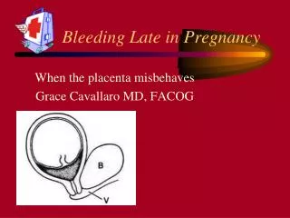
Placental Abruption • Premature separation of placenta from uterine wall • Partial or complete • “Marginal sinus separation” or “marginal sinus rupture” • Bleeding, but abnormal implantation or abruption never established
Epidemiology of Abruption • Occurs in 1-2% of pregnancies • Risk factors • hypertensive diseases of pregnancy • smoking or substance abuse (e.g. cocaine) • trauma • overdistension of the uterus • history of previous abruption • unexplained elevation of MSAFP • placental insufficiency • maternal thrombophilia/metabolic abnormalities
Abruption and Trauma • Can occur with blunt abdominal trauma and rapid deceleration without direct trauma • Complications inculde prematurity, growth restriction, stillbirth • Fetal evaluation after trauma • Increased use of FHR monitoring may decrease mortality
Bleeding from Abruption • Externalized hemorrhage • Bloody amniotic fluid • Retroplacental clot • 20% occult • “Couverlaire” uterus • Look for consumptive coagulopathy
Patient History - Abruption • Pain = hallmark symptom • Varies from mild cramping to severe pain • Back pain – think posterior abruption • Bleeding • May not reflect amount of blood loss • Differentiate from exuberant blood show • Trauma • Other risk factors (e.g. hypertension) • Membrane rupture
Physical Exam - Abruption • Signs of circulatory instability • Mild tachycardia normal • Signs and symptoms of shock represent > 30% blood test • Maternal abdomen • Fundal height • Leopold’s estimated fetal weight, fetal lie • Location of tenderness • Tetanic contractions
Ultrasound - Abruption • Abruption is a clinical diagnosis! • Placental location and appearance • Retroplacental echolucency • Abnormal thickening of placenta • “Torn” edge of placenta • Fetal lie • Estimated fetal weight
Laboratory - Abruption • Complete blood count • Type and Rh • Coagulation tests • Kleihauer-Betke not diagnostic, but useful to determine Rhogam dose • Preeclampsia labs, if indicated • Consider using drug screen
Treatment – Grade II Abruption • Assess fetal and maternal stability • Amniotomy • IUPC to detect elevated uterine tone • Expeditious operative or vaginal delivery • Maintain urine output > 30cc/hr and haematocrit > 30% • Prepare for neonatal resuscitation
Treatment – Grade III Abruption • Assess mother for hemodynamic and coagulation status • Vigorous replacement of fluid and blood products • Vaginal delivery preferred, unless severe haemorrhage
Coagulopathy with Abruption • Occurs in 1/3 of Grade III abruption • Usually not seen if live fetus • Etiologies: consumption, DIC • Administer platelets, FFP • Give factor VIII if severe
Epidemiology of Uterine Rupture • Occult dehiscence vs. symptomatic rupture • 0.03-0.08% of all women • 0.3-1.7% of women with uterine scar • Previous caesarean incision most common reason for scar disruption • Other causes: previous uterine curettage or perforation, inappropriate oxytocin usage, trauma
pervious uterine surgery congenital uterine anomaly uterine overdistension gestational trophoblastic neoplasia adenomyosis fetal anomaly vigorous uterine pressure difficult placental removal placenta increta or percreta Risk Factors – Uterine Rupture
Morbidity with Uterine Rupture • Maternal • haemorrhage with anaemia • bladder rupture • hysterectomy • maternal death • Fetal • respiratory distress • hypoxia • acidaemia • neonatal death
Patient History – Uterine Rupture • Vaginal bleeding • Pain • Cessation of contractions • Absence of FHR • Loss of station • Palpable fetal parts through maternal abdomen • Profound maternal tachycardia and hypotension
Uterine Rupture • Sudden deterioration of FHR pattern is most frequent finding • Placenta may play a role in uterine rupture • Transvaginal ultrasound to elevate uterine wall • MRI to confirm possible placenta accreta • Treatment • Asymptomatic scar disruption – expectant management • Symptomatic rupture – emergent caesarean delivery
Vasa Praevia • Rarest cause of haemorrhage • Onset with membrane rupture • Blood loss is fetal, with 50% mortality • Seen with low lying placenta, velamentous insertion of the cord or succenturiate lobe • Antepartum diagnosis • amnioscopy • colour doppler ultrasound • palpate vessels during vaginal examination
Diagnostic Tests – Vasa Praevia • Apt test – based on colorimetric response of fetal haemoglobin • Wright stain of vaginal bleed – for nucleated RBCs • Kleihauer-Betke test – 2 hour delay prohibits its use
Management – Vasa Praevia • Immediate caesarean delivery if fetal hear rate non-assuring • Administer normal saline 10-20 cc/kg bolus to newborn, if found to be in shock after delivery
Summary • Late pregnancy bleeding may herald diagnoses with significant morbidity/ mortality • Determining diagnosis important, as treatment dependent on cause • Avoid vaginal exam when placental location not known