Download
1 / 19
331 likes | 1.91k Vues
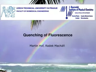
CZECH TECHNICAL UNIVERSITY IN PRAGUE FACULTY OF BIOMEDICAL ENGINEERING. Quenching of Fluorescence. Martin Hof, Radek Macháň. The fluorescence lifetime t = k -1 = ( k f + k nr ) -1 depends on the environment of the molecule through k nr = k i + k x + k ET + ….

E N D
CZECH TECHNICAL UNIVERSITY IN PRAGUE FACULTY OF BIOMEDICAL ENGINEERING Quenching of Fluorescence Martin Hof, Radek Macháň
The fluorescence lifetime t= k-1 = (kf + knr)-1 depends on the environment of the molecule through knr =ki + kx + kET + …. Fluorescence quantum yield: is proportional to fluorescence lifetime. Addition of another radiationless pathway increases knr and, thus, decreases t and QY. However, the measurement of fluorescence lifetime is more robust than measurement of fluorescence intensity (from which the QY is determined), because it depends on the intensity of excitation nor on the concentration of the fluorophores. The fluorescence intensityI (t) = kfn*(t) is proportional to n*(t) and vice versa
Quenching A number of processes can lead to a reduction in fluorescence intensity, i.e., quenching These processes can occur during the excited state lifetime – for example collisional quenching, energy transfer, charge transfer reactions or photochemistry – or they may occur due to formation of complexes in the ground state We shall focus our attention on the two quenching processes usually encountered – namely collisional(dynamic) quenching and static(complex formation) quenching Collisional Quenching Collisional quenching occurs when the excited fluorophore experiences contact with an atom or molecule that can facilitate non-radiative transitions to the ground state. Common quenchers include O2, I-, Cs+ and acrylamide. In the simplest case of collisional quenching, the following relation, called the Stern-Volmer equation, holds: I0/I = 1 + KSV[Q] where I0 and I are the fluorescence intensities observed in the absence and presence, respectively, of quencher, [Q] is the quencher concentration and KSV is the Stern-Volmer quenching constant
In the simplest case, then, a plot of I0/I versus [Q] should yield a straight line with a slope equal to KSV. Such a plot, known as a Stern-Volmer plot, is shown below for the case of fluorescein quenched by iodide ion (I-). In this case, KSV~ 8 Lmol-1 KSV = kq0 where kq is the bimolecular quenching rate constant and 0 is the excited state lifetime in the absence of quencher. I0/I In the case of purely collisional quenching, also known as dynamic quenching,: I0/I = 0/. Hence in this case: 0/ = 1 + kq 0[Q] In the fluorescein/iodide system, = 4ns and kq~ 2 x 10⁹ M-1 sec-1
Collisional Quenching derivation of Stern-Volmer equation: presence of quencher – additional nonradiative deexcitation channel quantum yield:
Static Quenching In some cases, the fluorophore can form a stable complex with another molecule. If this ground-state is non-fluorescent then we say that the fluorophore has been statically quenched. In such a case, the dependence of the fluorescence as a function of the quencher concentration follows the relation: I0/I = 1 + Ka[Q] where Kais the association constant of the complex. Such cases of quenching via complex formation were first described by Gregorio Weber. In the case of static quenching the lifetime of the sample will not be reduced since those fluorophores which are not complexed – and hence are able to emit after excitation – will have normal excited state properties. The fluorescence of the sample is reduced since the quencher is essentially reducing the number of fluorophores which can emit.
Static Quenching In some cases, the fluorophore can form a stable complex with another molecule. If this ground-state is non-fluorescent then we say that the fluorophore has been statically quenched. F + Q FQ [FQ] = Ka [F][Q]
I0/I [Q] If bothstaticanddynamicquenching are occurring in the sample then the following relation holds: I0/I = (1 + kq 0[Q]) (1 + Ka[Q]) In such a case then a plot of I0/I versus [Q] will have an upward curvature due to the [Q]2 term.
I0/I 0/ [Q] However, since the lifetime is unaffected by the presence of quencher in cases of pure static quenching, a plot of 0/ versus [Q] would give a straight line
I0/I [Q] Non-linear Stern-Volmer plots can also occur in the case of purely collisional quenching if some of the fluorophores are less accessible than others. Consider the case of multiple tryptophan residues in a protein – one can easily imagine that some of these residues would be more accessible to quenchers in the solvent than other. In the extreme case, a Stern-Volmer plot for a system having accessible and inaccessible fluorophores could look like this:
The quenching of LADH intrinsic protein fluorescence by iodide gives, in fact, just such a plot. LADH is a dimer with 2 tryptophan residues per identical monomer. One residue is buried in the protein interior and is relatively inaccessible to iodide while the other tryptophan residue is on the protein’s surface and is more accessible. 350nm 323nm E1 In this case (from Eftink and Selvidge, Biochemistry 1982, 21:117) the different emission wavelengths preferentially weigh the buried (323nm) or solvent exposed (350nm) tryptophan.
Laurdan DOPC DOPC DOTAP Patman E2 Where are the dyes localized? aqueous phase Laurdan and Patman are fluorescent probes which, thanks to their structure, incorporate to lipid bilayers (biological membranes) in well defined depths hydrophobic interior The depth can be determined by paralax method
Quenchers used for paralax method... E2 TEMPO-PC 5-DSA 16-DSA The quenchers used in paralax method are lipid analogues with groups with unpaired electrons at well defined positions (spin probes) – strong quenchers
Parallax method ... • Distance from the center of • DOPC bilayer for: • Patman – 10.45 A • Laurdan – 11.35 A E2
E3 Does the fluorophore change its location after addition of positively charged lipids? Patman Laurdan Quencher ... Acrylamide
E3 + + Stern-Volmer equation for dynamic quenching: Io/I= 1+KSV [Q] Positively charged dye (Patman) changes its location in the presence of positively charged lipids in the outward direction
Self quenching The intensity of fluorescence is proportional to the concentration of the fluorophores in a reasonable concentration range. However, at high concentrations of the fluorophores the proportionality is no more satisfied, because significant collisional quenching between the molecules of the fluorophore themselves appears. Fluorescence intensity of calcein as a function of fluorophore concentration Andersson et al. Eur Biophys J 2007, 36: 621
E4 Self quenching Typically used to study the formation of pores in vesicles caused by membrane-active molecules – vesicle leakage assay vesicles loaded with 60 mM calcein vesicles Triton X-100 detergent peptide LAH4 creates pores in the lipid bilayer, through which the dye can leak out LAH4 peptide detergent Triton X-100 micellizes the vesicles final calcein concentration ~ 5 mM Vogt and Bechinger, J Biol Chem 1999, 274: 29 115
Acknowledgement The course was inspired by courses of: Prof. David M. Jameson, Ph.D. Prof. RNDr. Jaromír Plášek, Csc. Prof. William Reusch Financial support from the grant: FRVŠ 33/119970