Download
1 / 12
150 likes | 422 Vues
Incomplete Ossification of the Humeral Condyle (IOHC) in Dogs. Bill Lee #103957. IOHC. “Max” Matthews eleven year old male German Shorthair Pointer fractured left humerus chasing a ball in the yard 103957. IOHC. “Max” Matthews Postop images 103957. IOHC.

E N D
Incomplete Ossification of the Humeral Condyle (IOHC) in Dogs Bill Lee #103957
IOHC • “Max” Matthews • eleven year old male German Shorthair Pointer • fractured left humerus chasing a ball in the yard • 103957
IOHC • “Max” Matthews • Postop images 103957
IOHC • IOHC described in 28 skeletally mature spaniels (Marcellin-Little, Vet. Surg. 1994) • Spaniels predisposed • Cocker, Brittany, Boykin, Springer, Clumber, Cavalier King Charles • Rottweiler, Labrador • Humeral condylar fractures (HCF) occur after minor trauma in these dogs
IOHC • Cause IOHC unknown: polygenic, recessive mode of inheritance • Some suggest elbow incongruity (reduced growth of distal ulnar physis) as causative • Male dogs predisposed, possibly b/c estrogen has stim. effect on epiphyseal maturation
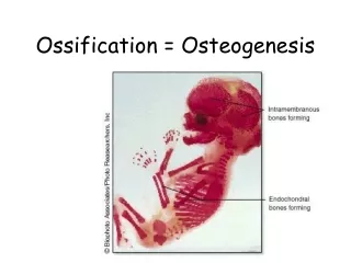
IOHC • Normal condylar ossification complete @ 8-12 weeks • 2 main oss. Centers (med. & lat. Aspect of condyle) fail to fuse • Remain separated by fibrous tiss.
IOHC • Classically, present with lameness or HCF at mean age = 6 yrs (range 2-12) • Some reports suggest obesity as a risk factor • All dogs w/ IOHC are bilaterally affected per Dr. Marcellin
Dx • Radiographic views: flexed L-M, hyperflexed L-M, Cr15°MCdLO • 5° off shown to obscure visualization • Radiolucent line on CrCd view, often approx. 1 mm wide, with surrounding sclerosis • Complete (spans articular surface to supratrochlear foramen) or incomplete
Dx • 92% of dogs with IOHC have concurrent DJD, 25% have FMCP • “Y” (93%), lat.(36%), med.(11%) fxs; lat. more common than med. (art. w/ radial head) • In imm. Dogs, IOHC cannot be confirmed. Must repeat rads after skeletal maturity
Dx • C.T. features of IOHC (Carrerra I, et. al. Vet. Surg. 2008.) • C.T. more sens. and spec. that rad. • MPR at the level of the medial coronoid apex (base and apex) • Joint space at humero-uln. art. wider than humero-rad. art. (incongruent) • Sig. diff. than normal elbows (apex > base)
Tx • Sx: transcond. lag screw, bone plates, etc • IOHC often does not heal after Sx, fibrous tiss. persists • Prophylactic transcond. Screw for IOHC w/out HCF appears to prevent lameness, fxs – no prospective studies