
LARGE INTESTINE
E N D
Presentation Transcript
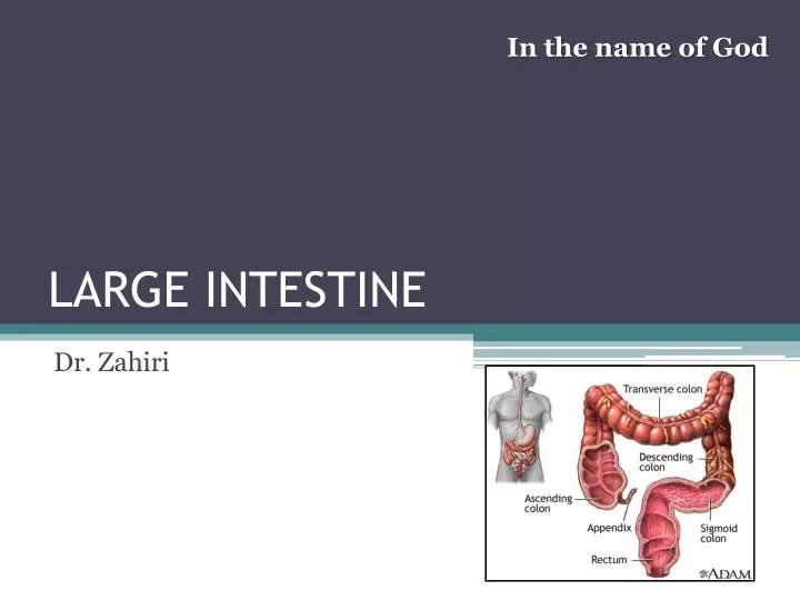
In the name of God LARGE INTESTINE Dr. Zahiri
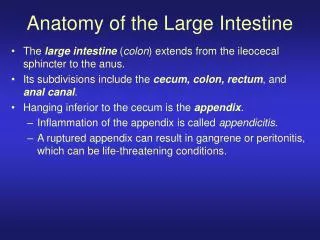
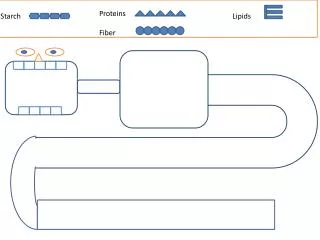
Dr. Maria Zahiri The large intestine (or colon) • the last part of the digestive system • Absorb water, sodium and some fat soluble vitamins. • The large intestine consists of : the cecum, appendix, colon, rectum, and anal canal. • is about 4.9 feet (1.5 m) long
Dr. Maria Zahiri Characteristics of large intestine • Taeniae coli: three bands of smooth muscle • Haustra : bulges caused by contraction of taeniae coli • Epiploic appendages (omental appendices ): are small pouches of the peritoneum filled with fat and situated along the colon. • their function is unknown.
Dr. Maria Zahiri Taeniae coli
Dr. Maria Zahiri Epiploic appendages
Dr. Maria Zahiri Caecum Position: Lie in the right iliac fossa below the ileocaecal valve. Is a intraperitoneal organ (?) Posteriorly lies the retrocaecal recess which frequently contains the vermiform appendix. Size:Average 6-7 cm
Dr. Maria Zahiri Blood supply
Dr. Maria Zahiri • Superior branch • Inferior branch • Appendicular artery • Ileal artery
Dr. Maria Zahiri Veins:
Dr. Maria Zahiri Lymphatic drainage • Anterior lymphatic vessels drain to: • Anterior ileocolicnodes • Posterior lymphatic vessels drains to: • Posterior ileocolic nodes • Inferior ileocolic nodes • To: Superior mesenteric nodes
Dr. Maria Zahiri Innervation: sympathetic and parasympathetic nerves via the superior mesenteric plexus.
Dr. Maria Zahiri Vermiform appendix
Dr. Maria Zahiri The vermiform appendix • is a narrow, vermian tube • arises from the posteromedial caecal wall • it varies from 5 to 20 cm in length, 2 cm below the end of the ileum.
Dr. Maria Zahiri The vermiform appendix It may occupy one of several positions: the commonest positions Retrocaecal(12 o’clock), retrocolic , pelvic or descending (4 o’clock) Other positions are occasionally seen especially when there is a long appendix mesentery allowing greater mobilit: subcaecal(6 o’clock); preilial(2 o’clock) ; postileal(2 o’clock) .
Dr. Maria Zahiri McBurney’s point
Dr. Maria Zahiri The three taeniae coli on the ascending colon and caecum converge on the base of the appendix, and merge into its longitudinal muscle. The anterior caecaltaeniais usually distinct and can be traced to the appendix. It is connected by a short mesoappendix to the lower part of the ileal mesentery.
Dr. Maria Zahiri The lumen of the appendix is small and opens into the caecum by an orifice lying below and slightly posterior to the ileocaecal opening. The orifice is sometimes guarded by a semi lunar mucosal fold forming a valve. The appendix usually contains numerous patches of lymphoid tissue although these tend to decrease in size from early adulthood.
Dr. Maria Zahiri VASCULAR SUPPLY • Ileocolic artery • Inferior branch • Appendicular artery • accessory arteries are common
Dr. Maria Zahiri Lymphatic vessels are numerous but all end in theinferiorandsuperior nodes of the ileocolic chain.
Dr. Maria Zahiri Innervation • sympathetic and parasympathetic nerves from the superior mesenteric plexus.
Dr. Maria Zahiri colon the colon consists of four sections: the ascending colon, the transverse colon, the descending colon, and the sigmoid colon
Dr. Maria Zahiri Ascending colon • narrower than the caecum- 15cm • It ascends to the inferior surface of the right lobe of the liver, on which it makes a shallow depression, and then turns abruptly forwards and to the left, at the hepatic flexure. • It is a retroperitoneal • Told fascia
Dr. Maria Zahiri TRANSVERS COLON • The transverse colon is 50 cm long • extends from the hepatic flexure in the right lumbar region across into the splenic flexure. • The transverse colon is suspended from the anterior border of the body of the pancreas by the transverse mesocolon.
Dr. Maria Zahiri SPLENIC FLEXURE • forms the junction of the transverse and descending colon • lies in the left hypochondriac region anteroinferior to the lower part of the spleen • The left kidney lies behind to it
Dr. Maria Zahiri • It lies more superiorly and posteriorly than the right hepatic flexure • is attached to the diaphragm at the level of the tenth and eleventh ribs by the phrenicocolic ligament which lies below the anterolateral pole of the spleen.
Dr. Maria Zahiri DESCENDING COLON • 25 cm • It descends through the left hypochondriac and lumbar regions • curves inferomedially to become the sigmoid colon at the inlet of the lesser pelvis. • It is a retroperitoneal structure covered anteriorly and on both sides by peritoneum.
Dr. Maria Zahiri SIGMOID COLON • begins at the pelvic inlet and ends at the rectum(S3) • It is completely invested in peritoneum and is attached to the posterior pelvic wall and lower posterior abdominal walls by the fan-shaped mesosigmoid. • The root of the sigmoid mesocolon has an inverted 'V‘ shape. • The position and shape of the sigmoid colon vary greatly
Dr. Maria Zahiri Left paracolic gutter Left ureter
Superior Mesenteric v. Middle colic a. Inf. pancresticodudenal a. Superior mesenteric a. Right colic a. Jejunal and ileal a. Ileocolic a. Appendicular a. Dr. Maria Zahiri
Inferior mesenteric v. Inferior mesenteric a. Left colic a. Sigmoid a. Superior rectal a. Dr. Maria Zahiri
Colic marginal artery Dr. Maria Zahiri
The Sudeck's point (or Sudeck's critical point): • refers to a specific location in the arterial supply of the rectosigmoid junction, namely the origin of the last sigmoid arterial branch from the inferior mesenteric artery (IMA) . • This arterial branch usually forms an anstomosis with a branch of the superior rectal artery. • The anastomosis is small and often only singular. Dr. Maria Zahiri
The "critical point" of Sudeck is marked with an "X." Dr. Maria Zahiri
Rectum Dr. Maria Zahiri • Rectum continuous with the sigmoid colon(S3) • Toupper end of the anal canal. Flexure: • the sacral flexure & the perinealflexure • three lateral curves : • upper is convex to the right, the middle (the most prominent) bulges to the left, and the lower is convex to the right • Both ends of the rectum are in the median plane
Dr. Maria Zahiri : 2/3anterior and 1/3lateral covered by peritoneum. the rectovesical pouch : The peritoneum is reflected superiorly onto the urinary bladder in males recto-uterine pouch (pouch of Douglas): • onto the posterior vaginal wall in females
ARTERIES Dr. Maria Zahiri 1.superior rectal artery: upper third 2.middle rectal artery:middle third (from Int. Iliac) 3.inferior rectal artery:distal third ( from Int. pudendal)
Dr. Maria Zahiri veins • 1.internal part: below the rectal and anal epithelium • 2.external part: outside the muscular wall
Dr. Maria Zahiri veins External plexus: • 1.inferior portion of the external plexus : • is drained by the inferior rectal vein into the internal pudendal vein • 2.middle portion : • by a middle rectal vein into the internal iliac vein, • 3.superior part : • By a superior rectal vein, which is the start of the inferior mesenteric vein. Communication between portal and systemic venous systems is thus established in the rectal plexus.
Dr. Maria Zahiri با آرزوی سلامت و سعادت