Download
1 / 45
450 likes | 488 Vues
This detailed review covers the myology of the wrist and hand, including bones, joints, ligaments, ranges of motion, and palpation techniques for students. Understand the intricate anatomy and functions of the wrist and hand for precise motor skills.

E N D
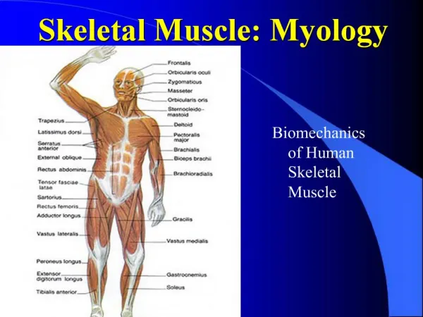
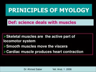
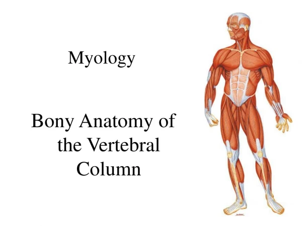
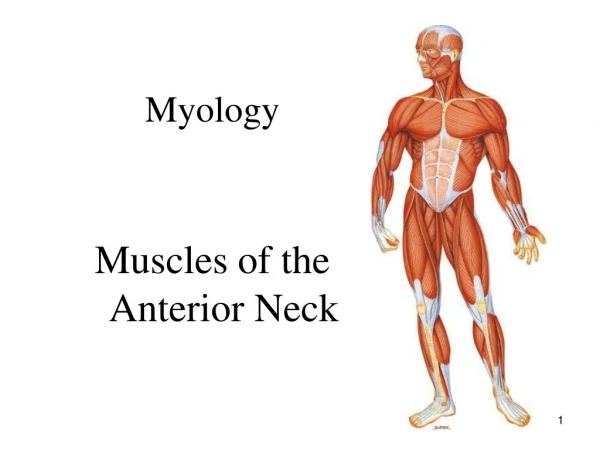
Myology Myology of the Wrist and Hand
Anatomical Review Distal Ulna and Radius (Notes in Lecture 3)
Carpals, Metacarpals, and Phalanges • Wrist: • Consists of eight small bones called carpals • Two rows • Proximal row: Scaphoid, Lunate, Triquetrum, and Pisiform • Distal row: Trapezium, Trapezoid, Capitate, Hamate • Palm: • Consists of the Metacarpals • Five metacarpals numbered I through V with number I being the thumb • Each metacarpal has a base, shaft, and a head • The base articulates with the carpals while the head articulates with the phalanges.
Fingers: • 14 phalanges in each hand (singular is phalanx) • Each phalanx also consists of a base, shaft, and head • The thumb only contains two phalanges, a proximal and distal one • The remaining fingers each contain three phalanges (proximal, middle, and distal) • Joints formed between phalanges: • Proximal interphalangeal joint (PIP) • Joint formed between proximal and middle phalanges • Distal interphalangeal joint (DIP) • Joint formed between middle and distal phalanges
Articular Anatomy of the Wrist The Wrist and hand are composed of 29 bones forming more than 20 joints. This allows us to perform fine delicate motor skills to forceful activity 2 joints make up the wrist: Radiocarpal: elliptical joint formed by the concavity of the radius and the convexity formed by the scaphoid and lunate. Ulnarcarpal joint: not a true articulation between the ulna and triquetrum. This articulation gives stability to the wrist. Movement within this joint has not been clearly defined.
Articular Anatomy of the Hand Four joints make up the hand: 1. Midcarpal joints: Gliding joints between carpal bones 2. Carpometacarpal joints: a. Carpometacarpal joint I: Rotated 90 degrees from metacarpal II. A Saddle joint allows for flexion/extension and abduction and adduction. b. Carpometacarpal joints II through IV: Gliding joints allowing slight sliding and flexion/extension. 3. Metocarpophalangeal joints: Ellipsoidal joints between the metacarpals and the proximal phalanges allowing for flexion/extension and abduction/adduction. 4. Interphalangeal joints: Hinge joints between the phalanges allowing for flexion and extension
Soft Tissue of the Wrist and Hand Radiocarpal Ligament: Connects the styloid process of the radius and distal aspect of the ulna to the scaphoid bone. Ulnar Collateral Ligament: attaches the styloid process of the ulna to the pisiform Radial Collateral Ligament: Attaches the styloid process of the radius to the radial side of the scaphoid and trapezium Flexor retinaculum: Wide thick ligament connecting the pisiform and hamate to the scaphoid and trapezium Palmar aponeurosis: formed from the deep fascia of the anterior hand, extending from the flexor retinaculum to the four fingers.
Ranges of Motion of the Wrist Each student should be able to describe and demonstrate the following movements: • Wrist flexion • Wrist extension • Abduction (Radial Deviation) • Adduction (Ulnar Deviation) • Wrist Circumduction
Ranges of Motion of the Fingers Each student should be able to describe and demonstrate the following movements: • Finger flexion • Finger extension • Finger abduction • Finger adduction • Finger circumduction
Ranges of Motion of the Thumb Each student should be able to describe and demonstrate the following movements: • Thumb flexion • Thumb extension • Thumb abduction • Thumb adduction • Thumb circumduction
Palpation of the wrist and fingers Carpals: Sitting; Locate the ulnar and radial styloid processes, slide your fingers distally, and then passively move the wrist in all directions and feel the carpals shift around a bit. Pisiform: Sitting; Locate the anterior wrist crease, move your fingers toward the pinky side, feel for the rounded shaped pisiform under the tissue of the palm. Hook of the Hamate: Sitting; Locate the pisiform again, draw an imaginary line from the pisiform to the index finger. Slide off the pisiform toward the index finger, about 3/4 of an inch from the pisiform feel for a small raised area. Metacarpals: Sitting; Palpate the bones of the palm both on the anterior and posterior surfaces. Feel the bases at the carpometacarpal joints, the shafts, and the enlarged heads at the metacarpophalangeal joints. Phalanges: Sitting; Locate the distal metacarpals again, then move distal palpating all three phalanges of the fingers (proximal, middle, and distal). Feel for the base, shafts, and heads of each phalange.
Anterior extrinsic muscles that move the wrist & hand Flexor Carpi Radialis Palmaris Longus Superficial layer Flexor Carpi Ulnaris Flexor Digitorum Superficialis Intermediate layer Flexor Digitorum Profundus Deep layer Flexor Pollicus Longus
O: Medial epicondyle (via common flexor tendon) of the humerus I: Base of the anterior aspect of the 2nd and 3rd metacarpals A: Flexion and radial deviation of the wrist. Assists in flexion and pronation of the forearm N: Median nerve Flexor Carpi Radialis
Palmaris Longus O: Medial epicondyle (via common flexor tendon) of the humerus I: Palmar aponeurosis of the hand A: Flexes the wrist. Assists in flexion of the elbow N: Median nerve
Flexor Carpi Ulnaris O: Humeral Head: Medial epicondyle (via common flexor tendon) of the humerus. Ulnar Head: olecranon process of the ulna I: Base of the 5th metacarpal, pisiform, and hook of hamate A: Flexion and ulnar deviation of the wrist. Assists in flexion of 3 the elbow. N: Ulnar
Flexor Digitorum Superficialis O: Humeroulnar Head: Medial epicondyle (via common flexor tendon) of the humerus. Radial Head: Anterior aspect of the mid-shaft of the radius I: Anterior surfaces of the middle phalanges of the 2nd through 5th digits at the PIP and MP’s. Assists in flexion of the elbow A: Flexion of the wrist and flexion of the 2nd through 5th digits N: Median nerve
Flexor Digitorum Profundus O: Mid shaft of the ulna and interosseous membrane I: anterior surface of the distal phalanges of 2nd through 5th digits A: Flexion of the wrist and flexion of the 2nd through 5th digits at the DIP. Assists in flexion of the wrist • Median and Ulnar nerves
Flexor Pollicus Longus O: Anterior aspect of the mid-shaft of the radius I: Distal phalanx of the thumb A: Flexes the wrist and flexion of the thumb N: Median nerve
Posterior extrinsic muscles that move the wrist and hand • Extensor Carpi Radialis Longus • Extensor Carpi Radialis Brevis • Extensor Carpi Ulnaris • Extensor Digitorum Communis Superficial Muscles • Extensor Digiti Minimi • Abductor Pollicis Longus • Extensor Pollicis Brevis • Extensor Pollicis Longus Deep Muscles • Extensor Indicis
Extensor Carpi Radialis Longus O: Lateral supracondylar ridge of the humerus I: Base of the posterior aspect of the 2nd metacarpal A: Extension and radial deviation of the wrist N: Radial nerve
Extensor Carpi Radialis Brevis • O: Lateral epicondyle of the humerus (via the common extensor tendon) • I: Base of the posterior • aspect of the 3rd metacarpal • A: Extension and radial • deviation of the wrist • N: Radial nerve
Extensor Carpi Ulnaris • O: Lateral epicondyle of the humerus (via the common extensor tendon) • I: Base of the posterior • aspect of the 5th metacarpal • A: Extension and ulnar deviation of the wrist • N: Radial nerve
Extensor Digitorum Communis • O: Lateral epicondyle of the humerus (via the common extensor tendon) • I: Phalanges of 2nd through the • 5th digits • A: Extension of the wrist and extension of the 2nd – 5th digits • N: Radial nerve
Extensor Digiti Minimi • O: Lateral epicondyle of the humerus (via the common extensor tendon) • I: Phalanx of the 5th digit • A: Extension of the pinky • N: Radial nerve
Abductor Pollicis Longus • O: Posterior radius and ulna • I: Base of the first metacarpal • A: Abduction of the thumb • N: Radial nerve
Extensor Pollicis Brevis • O: Posterior radius and • Interosseous membrane • I: Base of the phalanx of the thumb • A: Extension of the thumb • N: Radial nerve
Extensor Pollicis Longus O: Posterior ulna and Interosseous membrane I: Distal phalanx of the thumb A: Extension of the thumb N: Radial nerve
Extensor Indicis O: Posterior ulna and Interosseous membrane I: Index finger A: Extension of the index finger N: Radial nerve
Intrinsic Muscles of the Hand: Thenar Group Abductor pollicis brevis Opponens pollicis Flexor pollicis brevis Adductor pollicis All thenar muscles originate on carpal bones and insert into the thumb, hence they are agonists to thumb movement. All thenar muscles are innervated by the median nerve, with the exception of the adductor pollicis which innervated by the ulnar nerve.
Abductor pollicis brevis O: Scaphoid and trapezium I: Proximal phalanx of the thumb A: Abduction of the thumb N: Median nerve
Opponens pollicis O: Trapezium I: Lateral shaft of the 1st metacarpal A: Opposition of the thumb (flexion at MCP and phalangeal joint) N: Median nerve
Flexor pollicis brevis O: Trapezium I: Proximal phalanx of the thumb A: Flexion of the thumb N: Median nerve
Adductor pollicis O: Transverse Head: shaft of the 3rd metacarpal Oblique Head: carpals adjacent to the bases of the 2nd and 3rd metacarpals I: Base of the proximal phalanx of the thumb A: Adduction of the thumb N: Ulnar nerve
Intrinsic Muscles of the Hand: Hypothenar Group Palmaris Brevis Abductor Digiti Minimi Flexor Digiti Minimi Opponens Digiti Minimi All hypothenar muscles originate on the carpal bones and insert into the pinky All hypothenar muscles are innervated by the ulnar nerve
Palmaris Brevis O: Palmar aponeurosis I: Skin of the ulnar aspect of the hand A: Wrinkles skin of the palm N: Ulnar nerve Note: Palmaris brevis overlies the three other hypothenar muscles and is relatively unimportant, except that it covers and protects the ulnar nerve and artery
Abductor Digiti Minimi Manus O: Pisiform I: Proximal phalanx of the pinky A: Abducts the pinky N: Ulnar nerve
Flexor Digiti Minimi Manus O: Hook of hamate I: Proximal phalanx of the pinky A: Flexion of the pinky N: Ulnar nerve
Opponens Digiti Minimi O: Hook of hamate I: Ulnar border of the 5th metacarpal A: Opposition of the pinky N: Ulnar nerve
Intrinsic Muscles of the Hand: Mid-Palmar Group Lumbricales Manus Dorsal Interossei Manus Palmar Interossei Manus
Lumbricales Manus O: Distal tendons of the flexor digitorum profundus I: Extensor expansion of the 2nd through 5th fingers at proximal phalanges A: Flexion of the fingers at MCP joints and extension of the PIP and DIP’s N: Median nerve: 1st and 2nd lumbricales Ulnar nerve: 3rd and 4th lumbricales
Dorsal Interossei Manus O: Dorsal aspect of the metacarpals of the 2nd, 3rd, and 4th fingers I: Base of the proximal phalanges to extensor expansion of 2nd, 3rd, and 4th fingers A: Abduction of 2nd, 3rd, and 4th fingers (DAB). Dorsal interossei also assist Lumbricales in MCP flexion and PIP/DIP extension N: Ulnar nerve
Palmar Interossei Manus O: Palmar aspect of the 2nd, 4th, and 5th metacarpals I: Base of the proximal phalanges to extensor expansion of 2nd, 4th, and 5th fingers A: Adduction of 2nd, 4th, and 5th fingers (PAD). Palmar interossei also assist Lumbricales in MCP flexion and PIP/DIP extension N: Ulnar nerve