Anatomical Review: Myology of the Ankle & Foot Arches
260 likes | 332 Vues
Explore the intricacies of the ankle and foot myology, joints, ligaments, and ranges of motion. An in-depth study of tarsals, metatarsals, phalanges, foot arches, and articulations. Discover the articular anatomy and soft tissues of the ankle and foot.

Anatomical Review: Myology of the Ankle & Foot Arches
E N D
Presentation Transcript
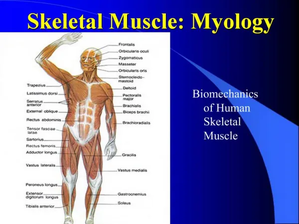
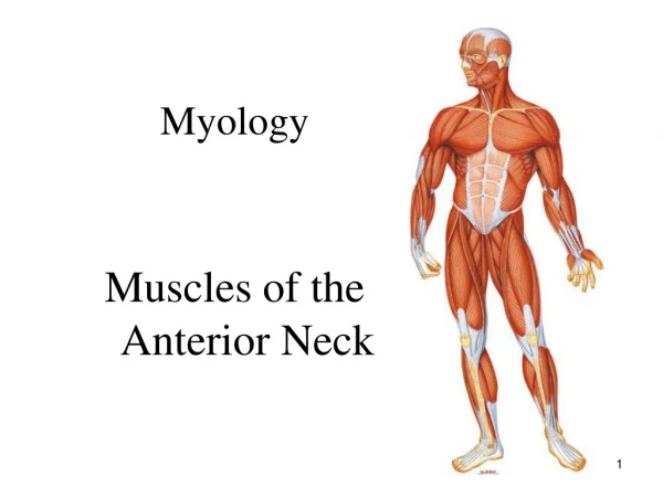
Myology Myology of the Ankle
Tarsals, Metatarsals, and Phalanges • Tarsals: • Consists of seven small bones called tarsals • Tarsals form the posterior half of the foot and heel. • Bones: Talus, calcaneus, cuboid, navicular, and three cuneiforms • Metatarsals • Same as the palm there are five metatarsals numbered I through V, with number I being the great toe • Each metatarsal has a base, shaft, and a head • The base articulates with the tarsals while the head articulates with the phalanges.
Toes: • 14 phalanges in each foot • Each phalanx also consists of a base, shaft, and head • The great toe only contains two phalanges, a proximal and distal one • The remaining toes each contain three phalanges (proximal, middle, and distal) • Joints formed between phalanges: • Proximal interphalangeal joint (PIP) • Joint formed between proximal and middle phalanges • Distal interphalangeal joint (DIP) • Joint formed between middle and distal phalanges
Arches of the Foot (See plate 5 for diagram of the arches of the foot) Functions: 1. Support the weight of the body 2. Provide an ideal distribution of the weight of the body over the hard and soft tissue of the foot. 3. Provide leverage while walking. • Arches are not rigid, they yield as weight is applied and spring back when weight is lifted thus helping absorb shock. • Arches develop about the age of 13 3 Arches: 1. Medial longitudinal arch: Originates at the calcaneus, rises to the talus, and descends through the navicular, the three cuneiforms, and the heads of the three medial metatarsals. 2. Lateral longitudinal arch: Originates at the calcaneus, rises to the cuboid, and descends to the heads of the lateral two metatarsals. 3. Transverse arch: Formed by the navicular, three cuneiforms, and bases of the five metatarsals.
Articular Anatomy: Ankle Ankle Joint: Also known as the talocrural joint • Formed by the articulation of: 1. Medial malleolus of the distal tibia and talus 2. Lateral malleolus of the distal fibula and talus • Considered to be a hinge joint Intertarsal joints: (3 articulations) 1. Subtalar joint: articulation between talus and calcaneus 2. Talocalcaneonavicular joint: articulation between talus, calcaneus, and navicular. 3. Calcaneocuboid joint: articulation between calcaneus and cuboid • These are considered to be planar (gliding) joints as the articular surfaces are flat or slightly curved.
Soft Tissue of the Ankle and Foot Articular Capsule: composed of fibrocartilage, thin anteriorly and posteriorly. Extends from the malleoli to the talus. Deltoid Ligament: Triangular shaped ligament on the medial aspect of the ankle. Apex is located at the medial malleolus. Base attaches to the talus, navicular, and calcaneus. Clinically, a very strong ligament. With eversion injuries the bones of the leg/ankle will most likely fracture before this ligament will tear. Plantar Calcaneonavicular (Spring) Ligament: Extends from the medial aspect of the calcaneus to the navicular.
Soft Tissue of the Ankle and Foot Lateral Ligaments: (considered some of the weakest ligaments in the body) a. Anterior Talofibular Ligament: Extends from the lateral malleolus to the talus b. Posterior Talofibular Ligament: Extends from the lateral malleolus to the talus c. Calcaneofibular Ligament: Extends from the lateral malleolus to the lateral aspect of the calcaneus.
Ankle and Foot Ranges of Motion Each student should be able to describe and demonstrate the following movements: • Ankle Dorsiflexion • Ankle Plantarflexion • Ankle Inversion • Ankle Eversion • Toe Flexion • Toe Extension • Toe Abduction • Toe Adduction • Hallux Flexion, Extension, Abduction, and Adduction Ankle joint Intertarsal joint Same as the fingers
Palpation of the Ankle and Foot Lateral and Medial Malleoli: Seated, palpate the large knobs on either side of the ankle. Note that the broader, medial malleolus is located at the distal end of the tibia while the more slender lateral malleolus is part of the fibula. Calcaneus: Seated, again locate the malleoli. Slide your fingers toward the heal and palpate the shape of the calcaneus. Cuneiforms: Seated, find the base of the 1st metatarsal. Then slide proximally into the “ditch” of the tarsometatarsal joint. Then continue proximally onto the surface of the medial cuneiform. To find the middle and lateral cuneiform, slide your fingers laterally along the dorsal surface of the foot. Navicular: Seated, again locate the base of the 1st metatarsal. Slide along the medial side of the foot, moving proximally across the surface of the medial cuneiform onto the navicular. Cuboid: Seated, locate the 5th metatarsal bone. Slide your finger along the 5th metatarsal to its base (which is expanded laterally). Continuing along the lateral side of the foot, slide your finger off the base of the 5th metatarsal onto the lateral border of the cuboid.
Muscles that move the ankle Tibialis Anterior Extensor Digitorum Longus Extensor Hallucis Longus Peroneus (Fibularis) Tertius Peroneus Longus Peroneus Brevis Gastrocnemius Soleus Plantaris Popliteus Flexor digitorum longus Flexor hallucis longus Tibialis posterior Anterior Compartment Lateral Compartment Posterior Compartment
Tibialis Anterior O: Entire shaft of the tibia I: 1st Cuneiform/1st Metatarsal A: Dorsiflexes and inversion of the ankle. N: Deep peroneal nerve Clinically, the Tibialis Anterior is related to shin splints, causing anterolateral leg pain
Extensor Digitorum Longus O: Proximal anterior fibula I: Middle and distal phalanges of the lateral four toes A: Extension of toes 2 through 5 and assists in dorsiflexion of the ankle. N: Deep peroneal nerve
Extensor Hallucis Longus O: Distal 1/3 of the anterior surface of the fibula and interosseous membrane I: Base of the distal phalange of the big toe. A: Extends the great toe and assists in dorsiflexion of the ankle. N: Deep peroneal nerve
Peroneus (Fibularis) Tertius O: Anterior surface of the distal fibula I: Base of the 5th metatarsal A: Dorsiflexion and eversion of the foot N: Deep peroneal nerve
Peroneus (Fibularis) Longus O: Proximal surface of the fibular head and shaft of the fibula I: Muscle cross under the foot from lateral to medial inserting onto the base of the 1st metatarsal and medial cuneiform. A: Eversion of the foot and assists in plantarflexion of the ankle. N: Superficial peroneal nerve
Peroneus (Fibularis) Brevis O: Lower 2/3 of lateral shaft of the fibula I: Base of the 5th metatarsal A: Eversion of the foot and assists in plantarflexion of the ankle. N: Superficial peroneal nerve
Gastrocnemius O: Posterior surface of the medial and lateral condyles I: Posterior surface of the calcaneus via the Achilles tendon. A. Knee: Flexion Ankle: Plantarflexion • Tibial nerve
Soleus O: Soleal line of tibia and fibular head. I: Calcaneus via the Achilles tendon. • Plantarflexion of the foot N: Tibial nerve
O: Lateral epicondyle of the femur I: Calcaneus via the Achilles tendon. A: Plantarflexion of the foot and flexion of the knee N: Tibial nerve Plantaris
O: Lateral condyle of the femur I: Posterior proximal shaft of the tibia A: Initiates knee flexion by medial rotation of the tibia to unlock the knee N: Tibial nerve Popliteus
O: Posterior surface of mid-shaft of the tibia I: Plantar surface of toes 2-5 A: Flexion of the toes, Plantarflexion and inversion of the ankle N: Tibial nerve Flexor Digitorum Longus
O: Posterior surface of fibula I: Plantar surface of the great toe A: Flexion of the great toe Plantarflexion and inversion of the ankle N: Tibial nerve Flexor Hallucis Longus
O: Posterior aspect of proximal tibia and fibula I: Plantar surface of the foot (navicular, adjacent tarsals, and metatarsals) A: Plantarflexion and inversion of the ankle N: Tibial nerve Tibialis Posterior