Cortex:
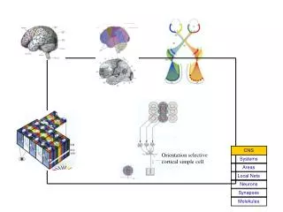
Orientation selective cortical simple cell. Cortex:. CNS. Systems. Areas . Local Nets. Neurons. Synapses. Molekules. Layers in the Cortex:. CNS. Systems. Areas . Local Nets. Neurons. Synapses. Molekules. CNS. Systems. Areas . Local Nets. Neurons. Synapses. Molekules.

Cortex:
E N D
Presentation Transcript
Orientation selective cortical simple cell Cortex: CNS Systems Areas Local Nets Neurons Synapses Molekules
Layers in the Cortex: CNS Systems Areas Local Nets Neurons Synapses Molekules
CNS Systems Areas Local Nets Neurons Synapses Molekules Local Circuits in V1: Circuit LGN inputs Cell types Spiny stellate cell Smooth stellate cell
Neurophysiological Background • “The Neuron “ • Contents: • Structure • Electrical Membrane Properties • Ion Channels • Actionpotential • Signal Propagation • Synaptic Transmission
CNS Systems Areas Local Nets Neurons Synapses Molekules Structure of a Neuron: At the dendrite the incoming signals arrive (incoming currents) At the soma current are finally integrated. At the axon hillock action potential are generated if the potential crosses the membrane threshold The axon transmits (transports) the action potential to distant sites At the synapses are the outgoing signals transmitted onto the dendrites of the target neurons
CNS Systems Areas Local Nets Neurons Synapses Molekules Structure of a Neuron: At the dendrite the incoming signals arrive (incoming currents) At the soma current are finally integrated. At the axon hillock action potential are generated if the potential crosses the membrane threshold The axon transmits (transports) the action potential to distant sites At the synapses are the outgoing signals transmitted onto the dendrites of the target neurons
Different Types of Neurons: dendrite dendrite Bipolar cell Unipolar cell axon soma axon soma Retinal bipolar cell (Invertebrate N.) Different Types of Multi-polar Cells Hippocampal pyramidal cell Purkinje cell of the cerebellum Spinal motoneuron
Cell membrane: The cell membrane separates intra- from extra-cellular spacesNa+ and Cl- ions are more concentrated outside, while negative ions (A-) and plenty of K+ are more concentrated inside. Due to differences in the ion-concenrations across the membrane a potential difference arises:In addition, the membrane acts like a capacitor:Current flow leads to voltage change: Cl- K+
rest rest Ion channels consist of big (protein) molecules which are inserted into to the membrane and connect intra- and extracellular space.Channels act as a restistance against the free flow of ions. Electrical resistor R:If Vm = Vrest there is no current flow.Channels are normally ion-selective and will open and close in dependence on the membrane potential (normal case) but also on (other) ions (e.g. NMDA channels). Channels exists for: K+, Na+, Ca2+, Cl- Ion channels:
rest rest rest In order to decribe the electrical properties of a membrane you need the membrane capacitance C, the conductivity g=1/R and the resting potential Vrest. In order to decribe the electrical properties of a membrane you need the membrane capacitance C, the conductivity g=1/R and the resting potential Vrest.Current across the membrane is given by: In order to decribe the electrical properties of a membrane you need the membrane capacitance C, the conductivity g=1/R and the resting potential Vrest.Current across the membrane is given by: or: In order to decribe the electrical properties of a membrane you need the membrane capacitance C, the conductivity g=1/R and the resting potential Vrest.Current across the membrane is given by: or:Using this equation you can calculate how the current changes depending on an experimentally injected current. Membrane - Circuit diagram:
Membrane - Circuit Diagram (advanced version): The whole thing gets more complicated due to the fact that there are many different ion channels all of which have their own characteristics depending on the momentarily existing state of the cell. The whole thing gets more complicated due to the fact that there are many different ion channels all of which have their own characteristics depending on the momentarily existing state of the cell. The conducitvity of a channel depends on the membrane potential and on the concentration difference between intra- and extracellular space (and sometimes also on other parameters). The whole thing gets more complicated due to the fact that there are many different ion channels all of which have their own characteristics depending on the momentarily existing state of the cell. The conducitvity of a channel depends on the membrane potential and on the concentration difference between intra- and extracellular space (and sometimes also on other parameters). One needs a computer simulation to describe this complex membrane behavior.
CNS Systems Areas Local Nets Neurons Synapses Molekules Structure of a Neuron: At the dendrite the incoming signals arrive (incoming currents). Signals propagate (normally) in a passive, electrotonic way towards the soma At the dendrite the incoming signals arrive (incoming currents). Signals propagate (normally) in a passive, electrotonic way towards the soma At the dendrite the incoming signals arrive (incoming currents). Signals propagate (normally) in a passive, electrotonic way towards the soma At the dendrite the incoming signals arrive (incoming currents). Signals propagate (normally) in a passive, electrotonic way towards the soma At the soma current are finally integrated. At the axon hillock action potential are generated if the potential crosses the membrane threshold The axon transmits (transports) the action potential to distant sites At the synapses are the outgoing signals transmitted onto the dendrites of the target neurons
Injected Current Membrane Potential Electrotonic Signal Propagation: Injected current flows out from the cell evenly across the membrane. Injected current flows out from the cell evenly across the membrane. The cell membrane has everywhere the same potential. Injected current flows out from the cell evenly across the membrane. The cell membrane has everywhere the same potential. The change in membrane potention follows an exponential with time constant: t = RC
The potential decays along a dendrite (or axon) according to the distance from the current injection site. The potential decays along a dendrite (or axon) according to the distance from the current injection site. At every location the temporal response follows an exponential but with ever decreasing amplitude. The potential decays along a dendrite (or axon) according to the distance from the current injection site. At every location the temporal response follows an exponential but with ever decreasing amplitude. If plotting only the maxima against the distance then you will get another exponential. The potential decays along a dendrite (or axon) according to the distance from the current injection site. At every location the temporal response follows an exponential but with ever decreasing amplitude. If plotting only the maxima against the distance then you will get another exponential. Different shape of the potentials in the dendrite and the soma of a motoneuron. Electrotonic Signal Propagation:
Compartment-Model: One can model the electrotonic propagation of potentials in the complex dendritic tree by subdividing the tree into small (cyklindrical) compartments. For each compartment the membrane equations can then be solved and integrated. (All this is tedious and complicated.)
CNS Systems Areas Local Nets Neurons Synapses Molekules Structure of a Neuron: At the dendrite the incoming signals arrive (incoming currents) At the soma current are finally integrated. At the axon hillock action potential are generated if the potential crosses the membrane threshold. At the axon hillock action potential are generated if the potential crosses the membrane threshold. At the axon hillock action potential are generated if the potential crosses the membrane threshold. At the axon hillock action potential are generated if the potential crosses the membrane threshold. The axon transmits (transports) the action potential to distant sites At the synapses are the outgoing signals transmitted onto the dendrites of the target neurons
Hodgkin Huxley Model: Na-Current + K-Current + Leakage Current + injec. Current
Hodgkin Huxley Model: + Leakage Current + injec. Strom + K-Current plus Equations for m and h
Hodgkin Huxley Model: + Leakage Current + injec. Current plus Equ. for m, h and n
Hodgkin Huxley Modell: rest plus Equ. For m, h and n
Hodgkin Huxley Modell: rest plus Equ. for m, h and n VNa= 55 mV, VK = -75 mV, Vrest = -60 mV gNa= 120 mS/cm2, gK= 36 mS/cm2, Cm= 1 mS/cm2
Iinj = 0.42 nA Short, weak current pulses depolarize the cell only a little. Iinj = 0.43 nA Iinj = 0.44 nA An action potential is elicited when crossing the threshold. Action Potential / Threshold:
Iinj = 0.45 nA A higher current reduces the time until an action potential is elicited. Iinj = 0.65 nA Iinj = 0.85 nA Action Potential / Firing Latency:
Action Potential / Refractory Period: Longer current pulses will lead to more action potentials. Longer current pulses will lead to more action potentials. However, directly after an action potential the ion channels are in an inactive state and cannot open. In addition, the membrane potential is rather hyperpolarized. Thus, the next action potential can only occur after a “waiting period” during which the cell return to its normal state. Longer current pulses will lead to more action potentials. However, directly after an action potential the ion channels are in an inactive state and cannot open. In addition, the membrane potential is rather hyperpolarized. Thus, the next action potential can only occur after a “waiting period” during which the cell return to its normal state. This “waiting period” is called the refractory period. Iinj = 0.5 nA Iinj = 0.5 nA Iinj = 0.5 nA
Action Potential / Firing Rate: Iinj = 0.2 nA When injecting current for longer durations an increase in current strength will lead to an increase of the number of action potentials per time. Thus, the firing rate of the neuron increases. When injecting current for longer durations an increase in current strength will lead to an increase of the number of action potentials per time. Thus, the firing rate of the neuron increases. The maximum firing rate is limited by the absolute refractory period. Iinj = 0.3 nA Iinj = 0.6 nA
Action Potential / Shapes: Cat - Heart Rat - Muscle Squid Giant Axon
CNS Systems Areas Local Nets Neurons Synapses Molekules Structure of a Neuron: At the dendrite the incoming signals arrive (incoming currents) At the soma current are finally integrated. At the axon hillock action potential are generated if the potential crosses the membrane threshold. The axon transmits (transports) the action potential to distant sites The axon transmits (transports) the action potential to distant sites The axon transmits (transports) the action potential to distant sites The axon transmits (transports) the action potential to distant sites At the synapses are the outgoing signals transmitted onto the dendrites of the target neurons
Open channels per mm2 membrane area Local current loops Time Distance Propagation of an Action Potential: Action potentials propagate without being diminished (active process). Action potentials propagate without being diminished (active process). All sites along a nerve fiber will be depolarized until the potential passes threshold. As soon as this happens a new AP will be elicited at some distance to the old one. Action potentials propagate without being diminished (active process). All sites along a nerve fiber will be depolarized until the potential passes threshold. As soon as this happens a new AP will be elicited at some distance to the old one. Main current flow is across the fiber.
Structure of a Neuron: At the dendrite the incoming signals arrive (incoming currents) At the soma current are finally integrated. At the axon hillock action potential are generated if the potential crosses the membrane threshold The axon transmits (transports) the action potential to distant sites CNS Systems At the synapses are the outgoing signals transmitted onto the dendrites of the target neurons At the synapses are the outgoing signals transmitted onto the dendrites of the target neurons At the synapses are the outgoing signals transmitted onto the dendrites of the target neurons At the synapses are the outgoing signals transmitted onto the dendrites of the target neurons Areas Local Nets Neurons Synapses Molekules
Chemical synapse Neurotransmitter Receptors
Neurotransmitters Chemicals (amino acids, peptides, monoamines) that transmit, amplify and modulate signals between neuron and another cell. Cause either excitatory or inhibitory PSPs. Glutamate – excitatory transmitter GABA, glycine – inhibitory transmitter
Synaptic Transmission: Synapses are used to transmit signals from the axon of a source to the dendrite of a target neuron. Synapses are used to transmit signals from the axon of a source to the dendrite of a target neuron. There are electrical (rare) and chemical synapses (very common) Synapses are used to transmit signals from the axon of a source to the dendrite of a target neuron. There are electrical (rare) and chemical synapses (very common) At an electrical synapse we have direct electrical coupling (e.g., heart muscle cells). Synapses are used to transmit signals from the axon of a source to the dendrite of a target neuron. There are electrical (rare) and chemical synapses (very common) At an electrical synapse we have direct electrical coupling (e.g., heart muscle cells). At a chemical synapse a chemical substance (transmitter) is used to transport the signal. Synapses are used to transmit signals from the axon of a source to the dendrite of a target neuron. There are electrical (rare) and chemical synapses (very common) At an electrical synapse we have direct electrical coupling (e.g., heart muscle cells). At a chemical synapse a chemical substance (transmitter) is used to transport the signal. Electrical synapses operate bi-directional and are extremely fast, chem. syn. operate uni-directional and are slower. Synapses are used to transmit signals from the axon of a source to the dendrite of a target neuron. There are electrical (rare) and chemical synapses (very common) At an electrical synapse we have direct electrical coupling (e.g., heart muscle cells). At a chemical synapse a chemical substance (transmitter) is used to transport the signal. Electrical synapses operate bi-directional and are extremely fast, chem. syn. operate uni-directional and are slower. Chemical synapses can be excitatory or inhibitory they can enhance or reduce the signal change their synaptic strength (this is what happens during learning).
Axon Structure of a Chemical Synapse: Motor Endplate (Frog muscle) Synaptic cleft vesicles Active zone Muscle fiber Presynaptic membrane Postsynaptic membrane Synaptic cleft
Pre-synaptic action potential Concentration of transmitter in the synaptic cleft Post-synaptic action potential What happens at a chemical synapse during signal transmission: The pre-synaptic action potential depolarises the axon terminals and Ca2+-channels open. The pre-synaptic action potential depolarises the axon terminals and Ca2+-channels open. Ca2+ enters the pre-synaptic cell by which the transmitter vesicles are forced to open and release the transmitter. The pre-synaptic action potential depolarises the axon terminals and Ca2+-channels open. Ca2+ enters the pre-synaptic cell by which the transmitter vesicles are forced to open and release the transmitter. Thereby the concentration of transmitter increases in the synaptic cleft and transmitter diffuses to the postsynaptic membrane. The pre-synaptic action potential depolarises the axon terminals and Ca2+-channels open. Ca2+ enters the pre-synaptic cell by which the transmitter vesicles are forced to open and release the transmitter. Thereby the concentration of transmitter increases in the synaptic cleft and transmitter diffuses to the postsynaptic membrane. Transmitter sensitive channels at the postsyaptic membrane open. Na+ and Ca2+ enter, K+ leaves the cell. An excitatory postsynaptic current (EPSC) is thereby generated which leads to an excitatory postsynaptic potential (EPSP).
Neurotransmitters and their (main) Actions: Transmitter Channel-typ Ion-current Action Transmitter Channel-typ Ion-current Action Acetylecholin nicotin. Receptor Na+ and K+ excitatory Transmitter Channel-typ Ion-current Action Acetylecholin nicotin. Receptor Na+ and K+ excitatory Glutamate AMPA / Kainate Na+ and K+ excitatory Transmitter Channel-typ Ion-current Action Acetylecholin nicotin. Receptor Na+ and K+ excitatory Glutamate AMPA / Kainate Na+ and K+ excitatory GABA GABAA-Receptor Cl- inhibitory Transmitter Channel-typ Ion-current Action Acetylecholin nicotin. Receptor Na+ and K+ excitatory Glutamate AMPA / Kainate Na+ and K+ excitatory GABA GABAA-Receptor Cl- inhibitory Glycine Cl- inhibitory Transmitter Channel-typ Ion-current Action Acetylecholin nicotin. Receptor Na+ and K+ excitatory Glutamate AMPA / Kainate Na+ and K+ excitatory GABA GABAA-Receptor Cl- inhibitory Glycine Cl- inhibitory Acetylecholin muscarin. Rec. - metabotropic, Ca2+ Release Transmitter Channel-typ Ion-current Action Acetylecholin nicotin. Receptor Na+ and K+ excitatory Glutamate AMPA / Kainate Na+ and K+ excitatory GABA GABAA-Receptor Cl- inhibitory Glycine Cl- inhibitory Acetylecholin muscarin. Rec. - metabotropic, Ca2+ Release Glutamate NMDA Na+, K+, Ca2+ voltage dependent blocked at resting potential
Long-term potentiation (LTP) High frequency stimulation: 1s, 100Hz
Long-term depression (LTD) Low frequency stimulation: 15min, 1Hz
Summation Properties at Synapses: Will be treated when we start to talk about: How to do calculations with neurons.
ui = signals from pre-synaptic neurons wi = synaptic weights S = Threshold(function) O = Output firing rate of the neuron The whole complex neuronal structure and function can be modeled at a first level of abstraction by a Simple Integrate-And-Fire Neuron: