Download
1 / 12
1.72k likes | 7.23k Vues
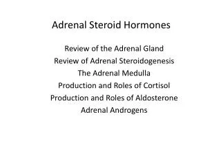
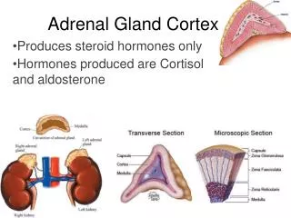
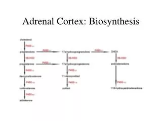
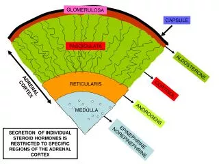
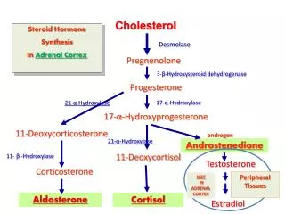
Cholesterol. Steroid Hormone Synthesis In Adrenal Cortex . Desmolase. Pregnenolone . 3- β - Hydroxysteroid dehydrogenase. Progesterone. 17- α -Hydroxylase. 21- α -Hydroxylase. 17- α - Hydroxyprogesterone. 11-Deoxycorticosterone. androgen. 21- α -Hydroxylase. Androstenedione .

E N D
Cholesterol Steroid Hormone Synthesis In Adrenal Cortex Desmolase Pregnenolone 3-β-Hydroxysteroid dehydrogenase Progesterone 17-α-Hydroxylase 21-α-Hydroxylase 17-α-Hydroxyprogesterone 11-Deoxycorticosterone androgen 21-α-Hydroxylase Androstenedione 11-Deoxycortisol 11- β-Hydroxylase Testosterone Corticosterone Peripheral Tissues NOT IN ADRENAL CORTEX Aldosterone Cortisol Estradiol
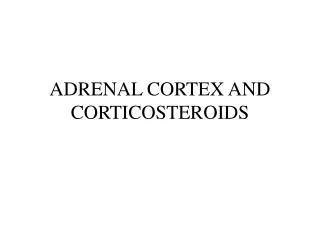
Pathway of Testosterone Production in the Testis Cholesterol In the Testis Androstenedione Testosterone 17b-hydroxysteroid Oxidoreductase N.B. In the adrenal cortex, androstendione(adrenal androgen) is formed. They are released to blood & converted in the testis (& peripheral tissues) to testosterone The production of androgens from cholesterol is identical to that in the adrenal, except that it continues from androstenedione to testosterone.
Pathway of Testosterone Production in the Testis • The main steroid produced in the male is testosteroneproduced from the testis. . • In the male, there is peripheral conversion of testosterone to: Dihydrotestosterone:in androgen target tissues, like muscles by 5 a reductase Or to • Estradiol : mostly in adipose tissue by enzyme cytochrome P450aromatase
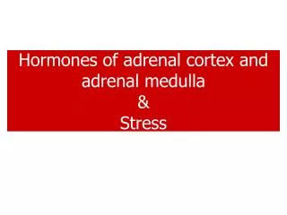
Synthesis of Steroid Hormones in the Ovary Stimulated by FSH Stimulated by LH Granulosa Cells of the Ovary • In the ovary: Estradiol is formed from the conversion of androgens (testosterone) into estradiol by the enzyme cytochrome P450 aromatase(in granulosa cells). The androgens required for conversion come from the neighboring theca cells. Cholesterol Theca Cells Androstendione Testosterone Estradiol • Aromatase
FSH LH LH receptor cholesterol Androstendione Estradiol Granulosa cells Theca cells aromatase AndrostendioneTestosterone Synthesis of steroid Hormones in the Ovary FSH ++ secretion of estrogen Regulates growth of ovarian follicle LH ++ estrogen secretion ++ ovulation FSH receptors LH & FSH stimulates estrogen secretion FSH regulates growth of ovarian follicles LH stimulates ovulation
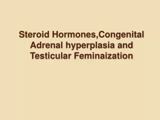
21 -Hydroxylase Deficiency 17-hydroxy-progesterone Progesterone Androstenedione In Peripheral Tissues 21 -hydroxylase 11-deoxycortisol 11-deoxycorticosterone Testosterone Cortisol Aldosterone Precocious sexual development in ♂ Virilisation of ♀
Case -1 : A 37 years old housewife Complaining of: Headache Weakness on trivial efforts Wasting in proximal limb muscles Polyuria Drinks water more than before On Examination: BP: 165/105 BMI: 33 Provisional Diagnosis: ?? Investigations: ?? Diagnosis: ??
Clinical Biochemistry Lab Investigations Fasting Blood Glucose: 160 mg/dl (N: 70 – 110 ) Urine glucose: nil Adrenal Function Tests Serum Cortisol at 8:00 AM : 410 nmol/L (N: 150 - 550) Serum Cortisol at 10:00 PM: 390 nmol/L (N: up to 200) Dexamethazone Suppression Test: Serum Cortisol: Basal (before dexamethazone 1 mg administration): 420 nmol/L After 48 hours of dexamethazone 1 mg administration: 410 nmol/L Insulin hypoglycemic Test: Serum Cortisol Basal at blood glucose 4.5 mmol/L: 435 nmol/L After blood glucose 1.5 mmol/L: 480 nmol/L ACTH at 8:00 AM: less than 2 ng/L (N: 7-51 )
Case -2: A 29-year-old female patient exhibited a rounded face, hirsutism, upper body obesity, easily bruised skin, severe fatigue, muscle weakness, and anxiety. She also complained of irregular periods. A long term sufferer, she had been prescribed prednisone for the past 2 years. Findings on examination revealed high fasting blood glucose levels and high blood pressure. Cortisol levels were below normal. Which one of the following is the most likely explanation to account for the patient’s symptoms? A. Decreased levels of insulin B. Increased levels of testosterone C. Decreased secretion of ACTH D. Excess exogenous glucocorticoid hormone E. Increased hepatic metabolism of steroid hormones
Prednisone acts as a glucocorticoid hormone analog, giving rise to Cushing syndrome symptoms after prolonged administration.
Case -3 A patient suffering from weakness, fatigue, nausea, and vomiting Low blood concentrations of Na+ and Cl− & high levels of serum K+. Physical examination revealed a deep tanning of both exposed and unexposed parts of the body and dark pigmentation inside the mouth. The hyperpigmentation in this patient most likely resulted from which of the following? A. Increased secretion of ACTH B. Prolonged exposure of the patient to ultraviolet radiation C. Excessive ingestion of β-carotene–containing foods D. Activation of melanocytes caused by medication side effects E. Inhibition of plasma membrane Na+, K+-ATPase
Hyperpigmentation is a feature of Addison disease, the diagnosis in this case. Decreased plasma cortisol because of adrenal insufficiency releases feedback increase of ACTH secretion by the pituitary, resulting in elevation of ACTH biosynthesis. The ACTH precursor peptide is cleaved to yield melanocyte-stimulating hormone (MSH)the factor responsible for hyperpigmentation even in areas not exposed to sunlight.